10
views
0
recommends
+1 Recommend
0 collections
    0
    shares
      • Record: found
      • Abstract: found
      • Article: found
      Is Open Access

      Breast cancer diagnosis based on lipid profiling by probe electrospray ionization mass spectrometry

      brief-report

      Read this article at

      Bookmark
          There is no author summary for this article yet. Authors can add summaries to their articles on ScienceOpen to make them more accessible to a non-specialist audience.

          Abstract

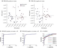
          Probe electrospray ionization mass spectrometry (PESI‐MS) is an ambient ionization‐based mass spectrometry method that surpasses the original electrospray ionization technique in features such as the rapidity of analysis, simplicity of the equipment and procedure, and lower cost. This study found that the PESI‐MS system with machine learning has the potential to establish a lipid‐based diagnosis of breast cancer with higher accuracy, using a simpler approach.

          Rapid mass spectrometry for breast cancer

          Related collections

          Most cited references17

          • Record: found
          • Abstract: found
          • Article: not found

          Machine Learning for Medical Imaging.

          Machine learning is a technique for recognizing patterns that can be applied to medical images. Although it is a powerful tool that can help in rendering medical diagnoses, it can be misapplied. Machine learning typically begins with the machine learning algorithm system computing the image features that are believed to be of importance in making the prediction or diagnosis of interest. The machine learning algorithm system then identifies the best combination of these image features for classifying the image or computing some metric for the given image region. There are several methods that can be used, each with different strengths and weaknesses. There are open-source versions of most of these machine learning methods that make them easy to try and apply to images. Several metrics for measuring the performance of an algorithm exist; however, one must be aware of the possible associated pitfalls that can result in misleading metrics. More recently, deep learning has started to be used; this method has the benefit that it does not require image feature identification and calculation as a first step; rather, features are identified as part of the learning process. Machine learning has been used in medical imaging and will have a greater influence in the future. Those working in medical imaging must be aware of how machine learning works. ©RSNA, 2017.
            Bookmark
            • Record: found
            • Abstract: found
            • Article: found

            Novel theranostic opportunities offered by characterization of altered membrane lipid metabolism in breast cancer progression.

            Activation of lipid metabolism is an early event in carcinogenesis and a central hallmark of many cancers. However, the precise molecular composition of lipids in tumors remains generally poorly characterized. The aim of the present study was to analyze the global lipid profiles of breast cancer, integrate the results to protein expression, and validate the findings by functional experiments. Comprehensive lipidomics was conducted in 267 human breast tissues using ultraperformance liquid chromatography/ mass spectrometry. The products of de novo fatty acid synthesis incorporated into membrane phospholipids, such as palmitate-containing phosphatidylcholines, were increased in tumors as compared with normal breast tissues. These lipids were associated with cancer progression and patient survival, as their concentration was highest in estrogen receptor-negative and grade 3 tumors. In silico transcriptomics database was utilized in investigating the expression of lipid metabolism related genes in breast cancer, and on the basis of these results, the expression of specific proteins was studied by immunohistochemistry. Immunohistochemical analyses showed that several genes regulating lipid metabolism were highly expressed in clinical breast cancer samples and supported also the lipidomics results. Gene silencing experiments with seven genes [ACACA (acetyl-CoA carboxylase α), ELOVL1 (elongation of very long chain fatty acid-like 1), FASN (fatty acid synthase), INSIG1 (insulin-induced gene 1), SCAP (sterol regulatory element-binding protein cleavage-activating protein), SCD (stearoyl-CoA desaturase), and THRSP (thyroid hormone-responsive protein)] indicated that silencing of multiple lipid metabolism-regulating genes reduced the lipidomic profiles and viability of the breast cancer cells. Taken together, our results imply that phospholipids may have diagnostic potential as well as that modulation of their metabolism may provide therapeutic opportunities in breast cancer treatment.
              Bookmark
              • Record: found
              • Abstract: found
              • Article: found
              Is Open Access

              Rapid evaporative ionisation mass spectrometry of electrosurgical vapours for the identification of breast pathology: towards an intelligent knife for breast cancer surgery

              Background Re-operation for positive resection margins following breast-conserving surgery occurs frequently (average = 20–25%), is cost-inefficient, and leads to physical and psychological morbidity. Current margin assessment techniques are slow and labour intensive. Rapid evaporative ionisation mass spectrometry (REIMS) rapidly identifies dissected tissues by determination of tissue structural lipid profiles through on-line chemical analysis of electrosurgical aerosol toward real-time margin assessment. Methods Electrosurgical aerosol produced from ex-vivo and in-vivo breast samples was aspirated into a mass spectrometer (MS) using a monopolar hand-piece. Tissue identification results obtained by multivariate statistical analysis of MS data were validated by histopathology. Ex-vivo classification models were constructed from a mass spectral database of normal and tumour breast samples. Univariate and tandem MS analysis of significant peaks was conducted to identify biochemical differences between normal and cancerous tissues. An ex-vivo classification model was used in combination with bespoke recognition software, as an intelligent knife (iKnife), to predict the diagnosis for an ex-vivo validation set. Intraoperative REIMS data were acquired during breast surgery and time-synchronized to operative videos. Results A classification model using histologically validated spectral data acquired from 932 sampling points in normal tissue and 226 in tumour tissue provided 93.4% sensitivity and 94.9% specificity. Tandem MS identified 63 phospholipids and 6 triglyceride species responsible for 24 spectral differences between tissue types. iKnife recognition accuracy with 260 newly acquired fresh and frozen breast tissue specimens (normal n = 161, tumour n = 99) provided sensitivity of 90.9% and specificity of 98.8%. The ex-vivo and intra-operative method produced visually comparable high intensity spectra. iKnife interpretation of intra-operative electrosurgical vapours, including data acquisition and analysis was possible within a mean of 1.80 seconds (SD ±0.40). Conclusions The REIMS method has been optimised for real-time iKnife analysis of heterogeneous breast tissues based on subtle changes in lipid metabolism, and the results suggest spectral analysis is both accurate and rapid. Proof-of-concept data demonstrate the iKnife method is capable of online intraoperative data collection and analysis. Further validation studies are required to determine the accuracy of intra-operative REIMS for oncological margin assessment. Electronic supplementary material The online version of this article (doi:10.1186/s13058-017-0845-2) contains supplementary material, which is available to authorized users.
                Bookmark

                Author and article information

                Contributors
                stakeda@yamanashi.ac.jp
                Journal
                Br J Surg
                Br J Surg
                10.1002/(ISSN)1365-2168
                BJS
                The British Journal of Surgery
                John Wiley & Sons, Ltd. (Chichester, UK )
                0007-1323
                1365-2168
                04 April 2020
                May 2020
                : 107
                : 6 ( doiID: 10.1002/bjs.v107.6 )
                : 632-635
                Affiliations
                [ 1 ] Department of Anatomy and Cell Biology, Faculty of Medicine Yamanashi Japan
                [ 2 ] Department of Digestive , Breast and Endocrine Surgery Yamanashi Japan
                [ 3 ] Department of Pathology, University of Yamanashi, Chu Yamanashi Japan
                [ 4 ] Shimadzu Corporation, Nakagyo Kyoto Japan
                Author notes
                [*] [* ] Correspondence to: Dr S. Takeda, Department of Anatomy and Cell Biology, Faculty of Medicine, University of Yamanashi, 1110 Shimokato, Chuo, Yamanashi, 409‐3898, Japan (e‐mail: stakeda@ 123456yamanashi.ac.jp )
                Author information
                https://orcid.org/0000-0002-4948-0389
                Article
                BJS11613
                10.1002/bjs.11613
                7216899
                32246473
                d002a326-2312-43b1-ab3d-46a26cd5056e
                © 2020 The Authors. British Journal of Surgery published by John Wiley & Sons Ltd on behalf of BJS Society Ltd.

                This is an open access article under the terms of the http://creativecommons.org/licenses/by-nc-nd/4.0/ License, which permits use and distribution in any medium, provided the original work is properly cited, the use is non?commercial and no modifications or adaptations are made.

                History
                : 17 February 2020
                : 08 March 2020
                Page count
                Figures: 2, Tables: 0, Pages: 4, Words: 2104
                Funding
                Funded by: Shimadzu corporation
                Categories
                Breast
                Rapid Research Communication
                Rapid Research Communication
                Custom metadata
                2.0
                May 2020
                Converter:WILEY_ML3GV2_TO_JATSPMC version:5.8.1 mode:remove_FC converted:12.05.2020

                Surgery
                Surgery

                Comments

                Comment on this article