723
views
0
recommends
+1 Recommend
0 collections
    15
    shares
      • Record: found
      • Abstract: found
      • Article: not found

      Oxidative stress and nerve damage: Role in chemotherapy induced peripheral neuropathy

      review-article

      Read this article at

      ScienceOpenPublisherPMC
      Bookmark
          There is no author summary for this article yet. Authors can add summaries to their articles on ScienceOpen to make them more accessible to a non-specialist audience.

          Abstract

          Peripheral neuropathy is a severe dose limiting toxicity associated with cancer chemotherapy. Ever since it was identified, the clear pathological mechanisms underlying chemotherapy induced peripheral neuropathy (CIPN) remain sparse and considerable involvement of oxidative stress and neuroinflammation has been realized recently. Despite the empirical use of antioxidants in the therapy of CIPN, the oxidative stress mediated neuronal damage in peripheral neuropathy is still debatable. The current review focuses on nerve damage due to oxidative stress and mitochondrial dysfunction as key pathogenic mechanisms involved in CIPN. Oxidative stress as a central mediator of apoptosis, neuroinflammation, metabolic disturbances and bioenergetic failure in neurons has been highlighted in this review along with a summary of research on dietary antioxidants and other nutraceuticals which have undergone prospective controlled clinical trials in patients undergoing chemotherapy.

          Graphical abstract

          Targeting chemotherpay induced peripheral neuropathy with natural antioxidants.

          Highlights

          • Oxidative stress contributes to the pathophysiology of chemotherapy induced peripheral neuropathies (CIPN).

          • Mitotoxicity and mitochondrial dysfunction contribute to amplified oxidative stress.

          • Pharmacological interventions targeted at maintenance of mitochondrial health and function may be beneficial against CIPN.

          Related collections

          Most cited references41

          • Record: found
          • Abstract: found
          • Article: not found

          Studies of peripheral sensory nerves in paclitaxel-induced painful peripheral neuropathy: evidence for mitochondrial dysfunction.

          Paclitaxel chemotherapy frequently induces neuropathic pain during and often persisting after therapy. The mechanisms responsible for this pain are unknown. Using a rat model of paclitaxel-induced painful peripheral neuropathy, we have performed studies to search for peripheral nerve pathology. Paclitaxel-induced mechano-allodynia and mechano-hyperalgesia were evident after a short delay, peaked at day 27 and finally resolved on day 155. Paclitaxel- and vehicle-treated rats were perfused on days 7, 27 and 160. Portions of saphenous nerves were processed for electron microscopy. There was no evidence of paclitaxel-induced degeneration or regeneration as myelin structure was normal and the number/density of myelinated axons and C-fibres was unaltered by paclitaxel treatment at any time point. In addition, the prevalence of ATF3-positive dorsal root ganglia cells was normal in paclitaxel-treated animals. With one exception, at day 160 in myelinated axons, total microtubule densities were also unaffected by paclitaxel both in C-fibres and myelinated axons. C-fibres were significantly swollen following paclitaxel at days 7 and 27 compared to vehicle. The most striking finding was significant increases in the prevalence of atypical (swollen and vacuolated) mitochondria in both C-fibres (1.6- to 2.3-fold) and myelinated axons (2.4- to 2.6-fold) of paclitaxel-treated nerves at days 7 and 27. Comparable to the pain behaviour, these mitochondrial changes had resolved by day 160. Our data do not support a causal role for axonal degeneration or dysfunction of axonal microtubules in paclitaxel-induced pain. Instead, our data suggest that a paclitaxel-induced abnormality in axonal mitochondria of sensory nerves contributes to paclitaxel-induced pain.
            Bookmark
            • Record: found
            • Abstract: found
            • Article: not found

            Chemotherapy-induced peripheral neuropathy: prevention and treatment strategies.

            Chemotherapy-induced peripheral neuropathy (CIPN) is a major dose limiting side effect of many commonly used chemotherapeutic agents, including platinum drugs, taxanes, epothilones and vinca alkaloids, and also newer agents such as bortezomib and lenolidamide. Symptom control studies have been conducted looking at ways to prevent or alleviate established CIPN. This manuscript provides a review of studies directed at both of these areas. New evidence strongly suggests that intravenous calcium and magnesium therapy can attenuate the development of oxaliplatin-induced CIPN, without reducing treatment response. Accumulating data suggest that vitamin E may also attenuate the development of CIPN, but more data regarding its efficacy and safety should be obtained prior to its general use in patients. Other agents that look promising in preliminary studies, but need substantiation, include glutamine, glutathione, N-acetylcysteine, oxcarbazepine, and xaliproden. Effective treatment of established CIPN, however, has yet to be found. Lastly, paclitaxel causes a unique acute pain syndrome which has been hypothesised to be caused by neurologic injury. No drugs, to date, have been proven to prevent this toxicity.
              Bookmark
              • Record: found
              • Abstract: found
              • Article: found
              Is Open Access

              Potential therapeutic effects of the simultaneous targeting of the Nrf2 and NF-κB pathways in diabetic neuropathy☆

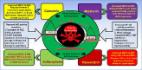
              Introduction Diabetes is one of the most debilitating conditions in patients affecting a substantial proportion of the world's population. Diabetes can predispose an individual to metabolic, cardiovascular disturbances and obesity, and these pathologies are accompanied by vascular complications [1]. Hyperglycaemia-induced damage to the endothelial cells results in micro-vascular complications of the diabetes such as diabetic neuropathy, nephropathy and retinopathy and macro-vascular complications such as cardiomyopathy [2]. Diabetic neuropathy remains the most severe form of complication affecting 40–50% of people with both types of diabetes. The clinical features of diabetic neuropathy range from sensory deficit to allodynia and hyperalgesia. Diabetic neuropathy arises from the long term effects of hyperglycaemia induced damage to peripheral nervous tissue as well as the vasa nervorum [3]. The current knowledge of pathophysiological mechanisms of hyperglycaemia-induced diabetic neuropathy is substantial and recent advances made in field could lead to the development of some novel therapeutic strategies targeted at advance glycation end products (AGE), sorbitol accumulation, protein kinase C (PKC) activation and hexosamine pathway. The axis of pathophysiological factors responsible for diabetes and diabetic neuropathy converge at two of the most extensively studied pathways, oxidative–nitrosative stress and neuroinflammation (Fig. 1). Molecular studies have revealed the involvement of transcriptional regulators such as Nrf2-Keap1 and the NF-κB inflammatory cascade in the pathophysiology of many diseases [4]. NF-κB has been shown to respond to the cellular redox status since a reducing environment prevents its activation whereas oxidative/nitrosative stress promotes phosphorylation and degradation of IκB [5]. Nrf2 increases intracellular GSH levels and GSH-dependent enzymes favouring a reducing environment thereby inhibiting NF-κB. Li et al. demonstrated that Nrf2-deficient mice exhibit greater induction of pro-inflammatory genes regulated by NF-κB such as interleukins, TNF-α, iNOS and COX-2 pointing towards the fact that Nrf2 deficiency enhances NF-κB-mediated pro-inflammatory reactions [6]. Soares et al. showed that HO-1 inhibited the TNF-α dependent activation of NF-κB in endothelial cells. It has been postulated that HO-1 induced by the Nrf2-EpRE interaction inhibits the NF-κB dependent transcriptional apparatus. Inhibition of NF-κB downstream of IκB phosphorylation/degradation and nuclear translocation has been hypothesized to be the site of action of HO-1 [11]. These data further support the concept that the Nrf2 directed increase in the expression of HO-1 is one of the hubs for cross-talk between Nrf2 and NF-κB (Figs. 2 and 3). Recent studies have shown that NF-κB suppresses the transcriptional activity of Nrf2. Liu et al. demonstrated that NF-κB p65 subunit repressed the beneficial effects of Nrf2 by promoting the localisation of transcription repressors, histone deacetylases with Nrf2/ARE and sequestering coactivators like CREB binding protein (CBP) [12]. Cells over-expressing NF-κB showed lesser expression of HO-1 which further confirms that NF-κB activation can act as a repressor of Nrf2 transcriptional activity. In a recent study, Yu et al. found that the N-terminal region of p65 subunit of NF-κB was physically associated with Keap1, and thus provide an additional mechanism for Nrf2–ARE inhibition. It was also suggested that NF-κB not only interacted with cytosolic Keap1 but also promoted nuclear translocation of Keap1 [13]. Previous studies with agents like curcumin [17], melatonin [18], resveratrol [19] and sulphoraphane [20] have reported beneficial effects in ameliorating various functional (motor nerve conduction velocity and nerve blood flow), sensorimotor (thermal and mechanical hyperalgesia) and biochemical deficits in experimental diabetic neuropathy (Fig. 4). These agents also suppressed the increased activity and levels of NF-κB and associated proteins and hence protected against neuroinflammation in diabetic neuropathy. As expected, treatment with these agents increased the levels of Nrf2 and HO-1 which further modulating the redox regulation of pro-inflammatory signalling pathways. Additional studies to find any common co-activators or co-repressors shared by these transcription factors and co-regulation by upstream and downstream signalling in these cascades will enable a better appreciation of the crosstalk between these two transcription factors in diabetic neuropathy. In summary, Nrf2 and NF-κB individually affect many signalling cascades to maintain a redox homeostasis; additionally they interact with each other to further modulate level of key redox modulators in health and disease. Studies with specific agents that might regulate the crosstalk between the two central pleiotropic transcription factors, Nrf2 and NF-κB, may be one of the prospective strategies that might aid in finding newer therapeutic choices for prevention and treatment of diabetic neuropathy.
                Bookmark

                Author and article information

                Journal
                Redox Biol
                Redox Biol
                Redox Biology
                Elsevier
                2213-2317
                18 January 2014
                18 January 2014
                2014
                : 2
                : 289-295
                Affiliations
                [0005]Department of Pharmacology and Toxicology, National Institute of Pharmaceutical Education and Research Hyderabad (NIPER-H), Bala Nagar, Hyderabad, AP 500037, India
                Author notes
                [* ]Corresponding author. Tel.: +91 40 23073741; fax: +91 40 23073751. ashutosh@ 123456niperhyd.ac.in ashutoshniper@ 123456gmail.com
                Article
                S2213-2317(14)00021-4
                10.1016/j.redox.2014.01.006
                3909836
                24494204
                d1a20a8b-b513-46df-949e-3d640c3860b3
                © 2014 The Authors
                History
                : 31 December 2013
                : 8 January 2014
                : 9 January 2014
                Categories
                Graphical Review

                peripheral neuropathy,chemotherapy,mitotoxicity,nutraceuticals,oxidative stress,mitochondria

                Comments

                Comment on this article