14
views
0
recommends
+1 Recommend
0 collections
    0
    shares
      • Record: found
      • Abstract: found
      • Article: found
      Is Open Access

      Contribution of epigenetic landscapes and transcription factors to X-chromosome reactivation in the inner cell mass

      research-article

      Read this article at

      Bookmark
          There is no author summary for this article yet. Authors can add summaries to their articles on ScienceOpen to make them more accessible to a non-specialist audience.

          Abstract

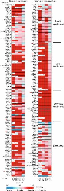
          X-chromosome inactivation is established during early development. In mice, transcriptional repression of the paternal X-chromosome (Xp) and enrichment in epigenetic marks such as H3K27me3 is achieved by the early blastocyst stage. X-chromosome inactivation is then reversed in the inner cell mass. The mechanisms underlying Xp reactivation remain enigmatic. Using in vivo single-cell approaches (allele-specific RNAseq, nascent RNA-fluorescent in situ hybridization and immunofluorescence), we show here that different genes are reactivated at different stages, with more slowly reactivated genes tending to be enriched in H3meK27. We further show that in UTX H3K27 histone demethylase mutant embryos, these genes are even more slowly reactivated, suggesting that these genes carry an epigenetic memory that may be actively lost. On the other hand, expression of rapidly reactivated genes may be driven by transcription factors. Thus, some X-linked genes have minimal epigenetic memory in the inner cell mass, whereas others may require active erasure of chromatin marks.

          Abstract

          X-chromosome inactivation is reversed in the mouse inner cell mass (ICM) through a mechanism that is not fully understood. Here, the authors investigate this process and characterize the contributions of the epigenetic landscape and transcription factors in X-linked gene reactivation dynamics.

          Related collections

          Most cited references41

          • Record: found
          • Abstract: found
          • Article: not found

          Gene Ontology: tool for the unification of biology

          Genomic sequencing has made it clear that a large fraction of the genes specifying the core biological functions are shared by all eukaryotes. Knowledge of the biological role of such shared proteins in one organism can often be transferred to other organisms. The goal of the Gene Ontology Consortium is to produce a dynamic, controlled vocabulary that can be applied to all eukaryotes even as knowledge of gene and protein roles in cells is accumulating and changing. To this end, three independent ontologies accessible on the World-Wide Web (http://www.geneontology.org) are being constructed: biological process, molecular function and cellular component.
            Bookmark
            • Record: found
            • Abstract: not found
            • Article: not found

            Gene action in the X-chromosome of the mouse (Mus musculus L.).

            MARY LYON (1961)
              Bookmark
              • Record: found
              • Abstract: found
              • Article: not found

              UTX and JMJD3 are histone H3K27 demethylases involved in HOX gene regulation and development.

              The trithorax and the polycomb group proteins are chromatin modifiers, which play a key role in the epigenetic regulation of development, differentiation and maintenance of cell fates. The polycomb repressive complex 2 (PRC2) mediates transcriptional repression by catalysing the di- and tri-methylation of Lys 27 on histone H3 (H3K27me2/me3). Owing to the essential role of the PRC2 complex in repressing a large number of genes involved in somatic processes, the H3K27me3 mark is associated with the unique epigenetic state of stem cells. The rapid decrease of the H3K27me3 mark during specific stages of embryogenesis and stem-cell differentiation indicates that histone demethylases specific for H3K27me3 may exist. Here we show that the human JmjC-domain-containing proteins UTX and JMJD3 demethylate tri-methylated Lys 27 on histone H3. Furthermore, we demonstrate that ectopic expression of JMJD3 leads to a strong decrease of H3K27me3 levels and causes delocalization of polycomb proteins in vivo. Consistent with the strong decrease in H3K27me3 levels associated with HOX genes during differentiation, we show that UTX directly binds to the HOXB1 locus and is required for its activation. Finally mutation of F18E9.5, a Caenorhabditis elegans JMJD3 orthologue, or inhibition of its expression, results in abnormal gonad development. Taken together, these results suggest that H3K27me3 demethylation regulated by UTX/JMJD3 proteins is essential for proper development. Moreover, the recent demonstration that UTX associates with the H3K4me3 histone methyltransferase MLL2 (ref. 8) supports a model in which the coordinated removal of repressive marks, polycomb group displacement, and deposition of activating marks are important for the stringent regulation of transcription during cellular differentiation.
                Bookmark

                Author and article information

                Contributors
                edith.heard@curie.fr
                Journal
                Nat Commun
                Nat Commun
                Nature Communications
                Nature Publishing Group UK (London )
                2041-1723
                3 November 2017
                3 November 2017
                2017
                : 8
                : 1297
                Affiliations
                [1 ]GRID grid.440907.e, Institut Curie, , PSL Research University, CNRS UMR3215, INSERM U934, UPMC Paris-Sorbonne, ; 26 Rue d’Ulm, 75005 Paris, France
                [2 ]ISNI 0000000121885934, GRID grid.5335.0, Department of Physiology, Development and Neuroscience, Wellcome Trust Cancer Research UK Gurdon Institute, , University of Cambridge, ; Tennis Court Road, Cambridge, CB2 1QN UK
                [3 ]ISNI 0000 0004 0372 2033, GRID grid.258799.8, Department of Anatomy and Cell Biology, Graduate School of Medicine, , Kyoto University, ; Yoshida-Konoe-cho, Sakyo-ku, Kyoto, 606-8501 Japan
                [4 ]ISNI 0000 0004 1754 9200, GRID grid.419082.6, JST, ERATO, ; Yoshida-Konoe-cho, Sakyo-ku, Kyoto, 606-8501 Japan
                [5 ]GRID grid.440907.e, Institut Curie, , PSL Research University, Mines Paris Tech, INSERM U900, ; 75005 Paris, France
                [6 ]ISNI 0000 0004 0605 769X, GRID grid.42475.30, Medical Research Council Laboratory of Molecular Biology, ; Francis Crick Avenue, Cambridge, CB2 0QH UK
                [7 ]ISNI 0000 0004 0372 2033, GRID grid.258799.8, Center for iPS Cell Research and Application, , Kyoto University, ; 53 Kawahara-cho, Shogoin, Sakyo-ku, Kyoto, 606-8507 Japan
                [8 ]ISNI 0000 0004 0372 2033, GRID grid.258799.8, Institute for Integrated Cell-Material Sciences, , Kyoto University, ; Yoshida-Ushinomiya-cho, Sakyo-ku, Kyoto, 606-8501 Japan
                [9 ]GRID grid.459340.f, Annoroad Gene Technology Co., Ltd, ; Beijing, 100176 China
                [10 ]ISNI 0000 0001 2111 7257, GRID grid.4488.0, Biotechnology Center, , Technische Universität Dresden, ; Tatzberg 47, 01307 Dresden, Germany
                Author information
                http://orcid.org/0000-0002-4378-5018
                http://orcid.org/0000-0002-2117-9754
                http://orcid.org/0000-0001-7319-043X
                http://orcid.org/0000-0003-2724-2002
                Article
                1415
                10.1038/s41467-017-01415-5
                5670228
                29101321
                d2dcc616-b184-4cfc-b9b4-9323791ce4b7
                © The Author(s) 2017

                Open Access This article is licensed under a Creative Commons Attribution 4.0 International License, which permits use, sharing, adaptation, distribution and reproduction in any medium or format, as long as you give appropriate credit to the original author(s) and the source, provide a link to the Creative Commons license, and indicate if changes were made. The images or other third party material in this article are included in the article’s Creative Commons license, unless indicated otherwise in a credit line to the material. If material is not included in the article’s Creative Commons license and your intended use is not permitted by statutory regulation or exceeds the permitted use, you will need to obtain permission directly from the copyright holder. To view a copy of this license, visit http://creativecommons.org/licenses/by/4.0/.

                History
                : 17 February 2017
                : 15 September 2017
                Categories
                Article
                Custom metadata
                © The Author(s) 2017

                Uncategorized
                Uncategorized

                Comments

                Comment on this article