23
views
0
recommends
+1 Recommend
0 collections
    0
    shares
      • Record: found
      • Abstract: found
      • Article: found
      Is Open Access

      Insulin and Glucagon Regulate Pancreatic α-Cell Proliferation

      research-article

      Read this article at

      Bookmark
          There is no author summary for this article yet. Authors can add summaries to their articles on ScienceOpen to make them more accessible to a non-specialist audience.

          Abstract

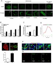
          Type 2 diabetes mellitus (T2DM) results from insulin resistance and β-cell dysfunction, in the setting of hyperglucagonemia. Glucagon is a 29 amino acid peptide hormone, which is secreted from pancreatic α cells: excessively high circulating levels of glucagon lead to excessive hepatic glucose output. We investigated if α-cell numbers increase in T2DM and what factor (s) regulate α-cell turnover. Lepr db/Lepr db (db/db) mice were used as a T2DM model and αTC1 cells were used to study potential α-cell trophic factors. Here, we demonstrate that in db/db mice α-cell number and plasma glucagon levels increased as diabetes progressed. Insulin treatment (EC50 = 2 nM) of α cells significantly increased α-cell proliferation in a concentration-dependent manner compared to non-insulin-treated α cells. Insulin up-regulated α-cell proliferation through the IR/IRS2/AKT/mTOR signaling pathway, and increased insulin-mediated proliferation was prevented by pretreatment with rapamycin, a specific mTOR inhibitor. GcgR antagonism resulted in reduced rates of cell proliferation in αTC1 cells. In addition, blockade of GcgRs in db/db mice improved glucose homeostasis, lessened α-cell proliferation, and increased intra-islet insulin content in β cells in db/db mice. These studies illustrate that pancreatic α-cell proliferation increases as diabetes develops, resulting in elevated plasma glucagon levels, and both insulin and glucagon are trophic factors to α-cells. Our current findings suggest that new therapeutic strategies for the treatment of T2DM may include targeting α cells and glucagon.

          Related collections

          Most cited references37

          • Record: found
          • Abstract: found
          • Article: not found

          SREBP Activity Is Regulated by mTORC1 and Contributes to Akt-Dependent Cell Growth

          Summary Cell growth (accumulation of mass) needs to be coordinated with metabolic processes that are required for the synthesis of macromolecules. The PI3-kinase/Akt signaling pathway induces cell growth via activation of complex 1 of the target of rapamycin (TORC1). Here we show that Akt-dependent lipogenesis requires mTORC1 activity. Furthermore, nuclear accumulation of the mature form of the sterol responsive element binding protein (SREBP1) and expression of SREBP target genes was blocked by the mTORC1 inhibitor rapamycin. We also show that silencing of SREBP blocks Akt-dependent lipogenesis and attenuates the increase in cell size in response to Akt activation in vitro. Silencing of dSREBP in flies caused a reduction in cell and organ size and blocked the induction of cell growth by dPI3K. Our results suggest that the PI3K/Akt/TOR pathway regulates protein and lipid biosynthesis in an orchestrated manner and that both processes are required for cell growth.
            Bookmark
            • Record: found
            • Abstract: found
            • Article: not found

            Selective versus total insulin resistance: a pathogenic paradox.

            Mice with type 2 diabetes manifest selective hepatic insulin resistance: insulin fails to suppress gluconeogenesis but continues to activate lipogenesis, producing the deadly combination of hyperglycemia and hypertriglyceridemia. In this issue of Cell Metabolism, Biddinger et al. (2008) show that mice with total hepatic insulin resistance exhibit hyperglycemia without hypertriglyceridemia-a state paradoxically less severe than selective insulin resistance.
              Bookmark
              • Record: found
              • Abstract: found
              • Article: not found

              Characterization of the cellular reduction of 3-(4,5-dimethylthiazol-2-yl)-2,5-diphenyltetrazolium bromide (MTT): subcellular localization, substrate dependence, and involvement of mitochondrial electron transport in MTT reduction.

              The MTT assay, which is widely used to measure cell proliferation and to screen for anticancer drugs, is based on reduction of the tetrazolium salt, MTT (3-(4,5-dimethylthiazol-2-yl)-2,5-diphenyltetrazolium bromide) by actively growing cells to produce a blue formazan product. Despite broad acceptance of this assay, neither the subcellular localization, nor the biochemical events involved in MTT reduction are known. Mitochondrial involvement in MTT reduction has been inferred from studies with respiratory inhibitors using succinate as a substrate, but the contribution of this activity to overall cellular MTT reduction is unknown. Using the bone marrow-derived cell line, 32D, we investigated the subcellular localization of MTT reduction using succinate, NADH, and NADPH as substrates. At optimum substrate concentrations, MTT reduction by whole cell homogenates was greatest with NADH and least with succinate, which accounted for less than 10% of the combined activities. Using succinate, 96% of recoverable MTT reducing activity was in particulate fractions of the cell and 77% in the mitochondrial and light mitochondrial/lysosomal fractions. When NADH and NADPH were used as substrates, increased amounts of MTT reducing activity were associated with soluble fractions of the cell and association with mitochondrial fractions was less pronounced. To further characterize MTT reduction by the mitochondrial fraction, respiratory chain inhibitors were used to explore involvement of electron transport in MTT reduction. Succinate-dependent mitochondrial MTT reduction was inhibited by 80% with chlorpromazine, 70% by antimycin A, and 85-90% by thenoyltrifluoracetone (TTFA), but inhibition was not observed with rotenone at < or = 2 microM, Amytal, or azide. These results suggest that when succinate is used as an electron donor, 70-80% of mitochondrial MTT reduction occurs subsequent to transfer of electrons from cytochrome c to cytochrome oxidase, but prior to the point of azide inhibition. In contrast to succinate, NADPH-dependent mitochondrial MTT reduction was not affected by any of the respiratory inhibitors tested, and NADH-dependent reduction was only inhibited by chlorpromazine (40-50% at plateau concentrations). These results suggest that most cellular MTT reduction occurs outside the mitochondrial inner membrane and involves NADH and NADPH-dependent mechanisms that are insensitive to respiratory chain inhibitors. This interpretation is supported by whole cell studies in which rotenone failed to affect basal and interleukin-3-stimulated MTT reduction at times up to 4 h but strongly inhibited DNA synthesis. We conclude that most cellular reduction of MTT occurs extramitochondrially and probably involves the pyridine nucleotide cofactors NADH and NADPH.
                Bookmark

                Author and article information

                Contributors
                Role: Editor
                Journal
                PLoS One
                plos
                plosone
                PLoS ONE
                Public Library of Science (San Francisco, USA )
                1932-6203
                2011
                25 January 2011
                : 6
                : 1
                : e16096
                Affiliations
                [1]National Institute on Aging, National Institutes of Health, Baltimore, Maryland, United States of America
                Mayo Clinic College of Medicine, United States
                Author notes

                Conceived and designed the experiments: ZL JME. Performed the experiments: ZL WK ZC YS ODC. Analyzed the data: ZL JME. Contributed reagents/materials/analysis tools: ZL WK ZC JLF LX JKN RS JOO QL. Wrote the paper: ZL JME.

                Article
                PONE-D-10-00682
                10.1371/journal.pone.0016096
                3026810
                21283589
                d2f43f61-33c7-4e53-970b-0b395f7de250
                This is an open-access article distributed under the terms of the Creative Commons Public Domain declaration which stipulates that, once placed in the public domain, this work may be freely reproduced, distributed, transmitted, modified, built upon, or otherwise used by anyone for any lawful purpose.
                History
                : 30 July 2010
                : 8 December 2010
                Page count
                Pages: 11
                Categories
                Research Article
                Biology
                Medicine
                Endocrinology

                Uncategorized
                Uncategorized

                Comments

                Comment on this article