34
views
0
recommends
+1 Recommend
0 collections
    0
    shares
      • Record: found
      • Abstract: found
      • Article: found
      Is Open Access

      Cabergoline, Dopamine D2 Receptor Agonist, Prevents Neuronal Cell Death under Oxidative Stress via Reducing Excitotoxicity

      research-article

      Read this article at

      Bookmark
          There is no author summary for this article yet. Authors can add summaries to their articles on ScienceOpen to make them more accessible to a non-specialist audience.

          Abstract

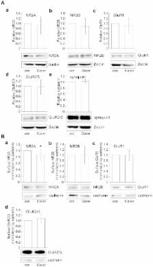
          Several lines of evidence demonstrate that oxidative stress is involved in the pathogenesis of neurodegenerative diseases, including Parkinson's disease. Potent antioxidants may therefore be effective in the treatment of such diseases. Cabergoline, a dopamine D2 receptor agonist and antiparkinson drug, has been studied using several cell types including mesencephalic neurons, and is recognized as a potent radical scavenger. Here, we examined whether cabergoline exerts neuroprotective effects against oxidative stress through a receptor-mediated mechanism in cultured cortical neurons. We found that neuronal death induced by H 2O 2 exposure was inhibited by pretreatment with cabergoline, while this protective effect was eliminated in the presence of a dopamine D 2 receptor inhibitor, spiperone. Activation of ERK1/2 by H 2O 2 was suppressed by cabergoline, and an ERK signaling pathway inhibitor, U0126, similarly protected cortical neurons from cell death. This suggested the ERK signaling pathway has a critical role in cabergoline-mediated neuroprotection. Furthermore, increased extracellular levels of glutamate induced by H 2O 2, which might contribute to ERK activation, were reduced by cabergoline, while inhibitors for NMDA receptor or L-type Ca 2+ channel demonstrated a survival effect against H 2O 2. Interestingly, we found that cabergoline increased expression levels of glutamate transporters such as EAAC1. Taken together, these results suggest that cabergoline has a protective effect on cortical neurons via a receptor-mediated mechanism including repression of ERK1/2 activation and extracellular glutamate accumulation induced by H 2O 2.

          Related collections

          Most cited references41

          • Record: found
          • Abstract: found
          • Article: not found

          Localization of neuronal and glial glutamate transporters.

          The cellular and subcellular distributions of the glutamate transporter subtypes EAAC1, GLT-1, and GLAST in the rat CNS were demonstrated using anti-peptide antibodies that recognize the C-terminal domains of each transporter. On immunoblots, the antibodies specifically recognize proteins of 65-73 kDa in total brain homogenates. Immunocytochemistry shows that glutamate transporter subtypes are distributed differentially within neurons and astroglia. EAAC1 is specific for certain neurons, such as large pyramidal cortical neurons and Purkinje cells, but does not appear to be selective for glutamatergic neurons. GLT-1 is localized only to astroglia. GLAST is found in both neurons and astroglia. The regional localizations are unique to each transporter subtype. EAAC1 is highly enriched in the cortex, hippocampus, and caudate-putamen and is confined to pre- and postsynaptic elements. GLT-1 is distributed in astrocytes throughout the brain and spinal cord. GLAST is most abundant in Bergmann glia in the cerebellar molecular layer brain, but is also present in the cortex, hippocampus, and deep cerebellar nuclei.
            Bookmark
            • Record: found
            • Abstract: found
            • Article: not found

            Neuroprotection by BDNF against glutamate-induced apoptotic cell death is mediated by ERK and PI3-kinase pathways.

            Neurotrophins protect neurons against glutamate excitotoxicity, but the signaling mechanisms have not been fully elucidated. We studied the role of the phosphatidylinositol 3-kinase (PI3-K) and Ras/mitogen-activated protein kinase (MAPK) pathways in the protection of cultured hippocampal neurons from glutamate induced apoptotic cell death, characterized by nuclear condensation and activation of caspase-3-like enzymes. Pre-incubation with the neurotrophin brain-derived neurotrophic factor (BDNF), for 24 h, reduced glutamate-evoked apoptotic morphology and caspase-3-like activity, and transiently increased the activity of the PI3-K and of the Ras/MAPK pathways. Inhibition of the PI3-K and of the Ras/MAPK signaling pathways abrogated the protective effect of BDNF against glutamate-induced neuronal death and similar effects were observed upon inhibition of protein synthesis. Moreover, incubation of hippocampal neurons with BDNF, for 24 h, increased Bcl-2 protein levels. The results indicate that the protective effect of BDNF in hippocampal neurons against glutamate toxicity is mediated by the PI3-K and the Ras/MAPK signaling pathways, and involves a long-term change in protein synthesis.
              Bookmark
              • Record: found
              • Abstract: found
              • Article: not found

              Mitochondrial dysfunction and oxidative stress in Parkinson's disease and monogenic parkinsonism.

              The pathogenic mechanisms that underlie Parkinson's disease remain unknown. Here, we review evidence from both sporadic and genetic forms of Parkinson's disease that implicate both mitochondria and oxidative stress as central players in disease pathogenesis. A systemic deficiency in complex I of the mitochondrial electron transport chain is evident in many patients with the disease. Oxidative stress caused by reactive metabolites of dopamine and alterations in the levels of iron and glutathione in the substantia nigra accompany this mitochondrial dysfunction. Recent evidence from studies on the genetic forms of parkinsonism with particular stress on DJ-1, parkin, and PINK-1 also suggest the involvement of mitochondria and oxidative stress. Copyright © 2012 Elsevier Inc. All rights reserved.
                Bookmark

                Author and article information

                Contributors
                Role: Editor
                Journal
                PLoS One
                PLoS ONE
                plos
                plosone
                PLoS ONE
                Public Library of Science (San Francisco, USA )
                1932-6203
                2014
                10 June 2014
                : 9
                : 6
                : e99271
                Affiliations
                [1 ]Department of Mental Disorder Research, National Institute of Neuroscience, National Center of Neurology and Psychiatry, Tokyo, Japan
                [2 ]Department of Life Science and Medical Bioscience, School of Advanced Science and Engineering, Waseda University, Tokyo, Japan
                [3 ]Core Research for Evolution Science and Technology Program (CREST), Japan Science and Technology Agency (JST), Tokyo, Japan
                Universidad de Castilla-La Mancha, Spain
                Author notes

                Competing Interests: The authors have declared that no competing interests exist.

                Conceived and designed the experiments: HO TN NA TI HK. Performed the experiments: HO TN YK NA. Analyzed the data: HO NA YO SN YK. Contributed reagents/materials/analysis tools: TN HK. Wrote the paper: HO TN.

                Article
                PONE-D-13-47278
                10.1371/journal.pone.0099271
                4051758
                24914776
                d31c5989-8888-4c7a-b9dd-197577257c08
                Copyright @ 2014

                This is an open-access article distributed under the terms of the Creative Commons Attribution License, which permits unrestricted use, distribution, and reproduction in any medium, provided the original author and source are credited.

                History
                : 12 November 2013
                : 13 May 2014
                Page count
                Pages: 12
                Funding
                This study was supported by the Intramural Research Grant (No. 24-11) for Neurological and Psychiatric Disorders of NCNP ( http://www.ncnp.go.jp/) and CREST program “creation of a novel technology towards diagnosis and treatment based on understanding of molecular pathogenesis of psychiatric and neurological disorders” (No. 08062571) of JST ( http://www.sss.jst.go.jp/info/index.html) (T.N., N.A. and H.K.). This project was also supported by Takeda Science Foundation ( http://www.takeda-sci.or.jp/) (T.N.), a grant from Grant-in-Aid for Scientific Research (B) (JSPS KAKENHI Grant Number 24300139) ( http://www.jsps.go.jp/english/index.html) (T.N.), and Grant-in-Aid for Challenging Exploratory Research (JSPS KAKENHI Grant Number 25640019) ( http://www.jsps.go.jp/english/index.html) (T.N.) from the Ministry of Education, Culture, Sports, Science, and Technology of Japan. The funders had no role in study design, data collection and analysis, decision to publish, or preparation of the manuscript.
                Categories
                Research Article
                Biology and life sciences
                Biochemistry
                Hormones
                Catecholamines
                Dopamine
                Neurochemistry
                Neurochemicals
                Cell biology
                Cell Processes
                Cell Death
                Cellular Types
                Animal Cells
                Neurons
                Signal transduction
                Cell signaling
                Signaling cascades
                ERK signaling cascade
                Molecular Cell Biology
                Toxicology
                Neurotoxicology
                Medicine and Health Sciences
                Clinical Medicine
                Pharmacology
                Drug Research and Development

                Uncategorized
                Uncategorized

                Comments

                Comment on this article