48
views
0
recommends
+1 Recommend
0 collections
    0
    shares
      • Record: found
      • Abstract: found
      • Article: found
      Is Open Access

      Characterisation of the active/de-active transition of mitochondrial complex I

      review-article
      , , , *
      Biochimica et Biophysica Acta
      Elsevier Pub. Co
      A/D, active/de-active transition, BN-PAGE, blue native polyacrylamide gel electrophoresis, DIGE, difference gel electrophoresis, DTNB, 5,5′-dithiobis-(2-nitrobenzoic acid), EEDQ, N-ethoxycarbonyl-2-ethoxy-1,2-dihydroquinoline, EMCS, N-ε-maleimidocaproyl-oxysuccinimide ester, GSH/GSSG, reduced/oxidised glutathione, HAR, hexaammineruthenium, hrCN-PAGE, high resolution clear native polyacrylamide gel electrophoresis, I/R, ischemia/reperfusion, NADH, dihydronicotinamide adenine dinucleotide, NEM, N-ethylmaleimide, NHS, N-hydroxysuccinimide, NO, nitric oxide, Q, ubiquinone, RNS, reactive nitrogen species, ROS, reactive oxygen species, SDS-PAGE, sodium dodecyl sulphate polyacrylamide gel electrophoresis, SMP, submitochondrial particles, SPDP, N-succinimidyl 3-(2-pyridyldithio)-propionate, Mitochondrial complex I, A/D transition, Conformational change, Ischaemia/reperfusion, Thiol modification

      Read this article at

      Bookmark
          There is no author summary for this article yet. Authors can add summaries to their articles on ScienceOpen to make them more accessible to a non-specialist audience.

          Abstract

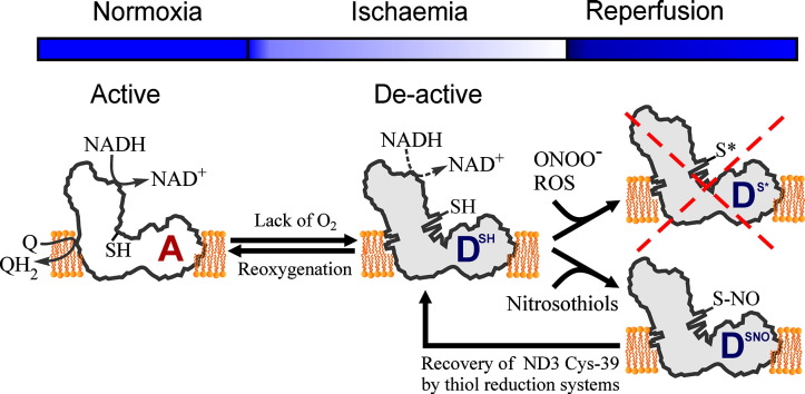
          Oxidation of NADH in the mitochondrial matrix of aerobic cells is catalysed by mitochondrial complex I. The regulation of this mitochondrial enzyme is not completely understood. An interesting characteristic of complex I from some organisms is the ability to adopt two distinct states: the so-called catalytically active (A) and the de-active, dormant state (D). The A-form in situ can undergo de-activation when the activity of the respiratory chain is limited (i.e. in the absence of oxygen).

          The mechanisms and driving force behind the A/D transition of the enzyme are currently unknown, but several subunits are most likely involved in the conformational rearrangements: the accessory subunit 39 kDa (NDUFA9) and the mitochondrially encoded subunits, ND3 and ND1. These three subunits are located in the region of the quinone binding site.

          The A/D transition could represent an intrinsic mechanism which provides a fast response of the mitochondrial respiratory chain to oxygen deprivation. The physiological role of the accumulation of the D-form in anoxia is most probably to protect mitochondria from ROS generation due to the rapid burst of respiration following reoxygenation. The de-activation rate varies in different tissues and can be modulated by the temperature, the presence of free fatty acids and divalent cations, the NAD +/NADH ratio in the matrix, the presence of nitric oxide and oxygen availability.

          Cysteine-39 of the ND3 subunit, exposed in the D-form, is susceptible to covalent modification by nitrosothiols, ROS and RNS. The D-form in situ could react with natural effectors in mitochondria or with pharmacological agents. Therefore the modulation of the re-activation rate of complex I could be a way to ameliorate the ischaemia/reperfusion damage. This article is part of a Special Issue entitled: 18th European Bioenergetic Conference. Guest Editors: Manuela Pereira and Miguel Teixeira.

          Graphical abstract

          Highlights

          • The potential mechanism of complex I A/D transition is discussed.

          • An —SH group exposed in the D-form is susceptible to covalent modification.

          • The role of A/D transition in tissue response to ischaemia is proposed.

          Related collections

          Most cited references137

          • Record: found
          • Abstract: found
          • Article: not found

          Supercomplexes in the respiratory chains of yeast and mammalian mitochondria.

          Around 30-40 years after the first isolation of the five complexes of oxidative phosphorylation from mammalian mitochondria, we present data that fundamentally change the paradigm of how the yeast and mammalian system of oxidative phosphorylation is organized. The complexes are not randomly distributed within the inner mitochondrial membrane, but assemble into supramolecular structures. We show that all cytochrome c oxidase (complex IV) of Saccharomyces cerevisiae is bound to cytochrome c reductase (complex III), which exists in three forms: the free dimer, and two supercomplexes comprising an additional one or two complex IV monomers. The distribution between these forms varies with growth conditions. In mammalian mitochondria, almost all complex I is assembled into supercomplexes comprising complexes I and III and up to four copies of complex IV, which guided us to present a model for a network of respiratory chain complexes: a 'respirasome'. A fraction of total bovine ATP synthase (complex V) was isolated in dimeric form, suggesting that a dimeric state is not limited to S.cerevisiae, but also exists in mammalian mitochondria.
            Bookmark
            • Record: found
            • Abstract: found
            • Article: not found

            Supercomplex assembly determines electron flux in the mitochondrial electron transport chain.

            The textbook description of mitochondrial respiratory complexes (RCs) views them as free-moving entities linked by the mobile carriers coenzyme Q (CoQ) and cytochrome c (cyt c). This model (known as the fluid model) is challenged by the proposal that all RCs except complex II can associate in supercomplexes (SCs). The proposed SCs are the respirasome (complexes I, III, and IV), complexes I and III, and complexes III and IV. The role of SCs is unclear, and their existence is debated. By genetic modulation of interactions between complexes I and III and III and IV, we show that these associations define dedicated CoQ and cyt c pools and that SC assembly is dynamic and organizes electron flux to optimize the use of available substrates.
              Bookmark
              • Record: found
              • Abstract: found
              • Article: not found

              Crystal structure of the entire respiratory complex I

              Complex I is the first and largest enzyme of the respiratory chain, playing a central role in cellular energy production by coupling electron transfer between NADH and ubiquinone to proton translocation. It is implicated in many common human neurodegenerative diseases. Here we report the first crystal structure of the entire, intact complex I (from T. thermophilus) at 3.3 Å resolution. The structure of the 536 kDa complex comprises 16 different subunits with 64 transmembrane helices and 9 Fe-S clusters. The core fold of subunit Nqo8 (NuoH/ND1) is, unexpectedly, similar to a half-channel of the antiporter-like subunits. Small subunits nearby form a linked second half-channel, thus completing the fourth proton translocation pathway, in addition to the channels in three antiporter-like subunits. The quinone-binding site is unusually long, narrow and enclosed. The quinone headgroup binds at the deep end of this chamber, near cluster N2. Strikingly, the chamber is linked to the fourth channel by a “funnel” of charged residues. The link continues over the entire membrane domain as a remarkable flexible central axis of charged and polar residues. It likely plays a leading role in the propagation of conformational changes, aided by coupling elements. The structure suggests that a unique, out-of-the-membrane quinone reaction chamber allows the redox energy to drive concerted long-range conformational changes in the four antiporter-like domains, resulting in translocation of four protons per cycle.
                Bookmark

                Author and article information

                Contributors
                Journal
                Biochim Biophys Acta
                Biochim. Biophys. Acta
                Biochimica et Biophysica Acta
                Elsevier Pub. Co
                0006-3002
                1 July 2014
                July 2014
                : 1837
                : 7
                : 1083-1092
                Affiliations
                Queen's University Belfast, School of Biological Sciences, Medical Biology Centre, 97 Lisburn Road, Belfast BT9 7BL, UK
                Author notes
                [* ]Corresponding author. Tel.: + 44 28 9097 2166; fax: + 44 28 9097 5877. a.galkin@ 123456qub.ac.uk
                Article
                S0005-2728(14)00065-6
                10.1016/j.bbabio.2014.02.018
                4331042
                24569053
                d5407b11-4f2b-431a-814f-26e6767264a4
                © 2014 The Authors

                This is an open access article under the CC BY license (http://creativecommons.org/licenses/by/3.0/).

                History
                : 30 November 2013
                : 14 February 2014
                : 17 February 2014
                Categories
                Review

                Biochemistry
                a/d, active/de-active transition,bn-page, blue native polyacrylamide gel electrophoresis,dige, difference gel electrophoresis,dtnb, 5,5′-dithiobis-(2-nitrobenzoic acid),eedq, n-ethoxycarbonyl-2-ethoxy-1,2-dihydroquinoline,emcs, n-ε-maleimidocaproyl-oxysuccinimide ester,gsh/gssg, reduced/oxidised glutathione,har, hexaammineruthenium,hrcn-page, high resolution clear native polyacrylamide gel electrophoresis,i/r, ischemia/reperfusion,nadh, dihydronicotinamide adenine dinucleotide,nem, n-ethylmaleimide,nhs, n-hydroxysuccinimide,no, nitric oxide,q, ubiquinone,rns, reactive nitrogen species,ros, reactive oxygen species,sds-page, sodium dodecyl sulphate polyacrylamide gel electrophoresis,smp, submitochondrial particles,spdp, n-succinimidyl 3-(2-pyridyldithio)-propionate,mitochondrial complex i,a/d transition,conformational change,ischaemia/reperfusion,thiol modification

                Comments

                Comment on this article