28
views
0
recommends
+1 Recommend
0 collections
    0
    shares
      • Record: found
      • Abstract: found
      • Article: found
      Is Open Access

      The Immunometabolomic Interface Receptor Hydroxycarboxylic Acid Receptor 2 Mediates the Therapeutic Effects of Dimethyl Fumarate in Autoantibody-Induced Skin Inflammation

      research-article

      Read this article at

      Bookmark
          There is no author summary for this article yet. Authors can add summaries to their articles on ScienceOpen to make them more accessible to a non-specialist audience.

          Abstract

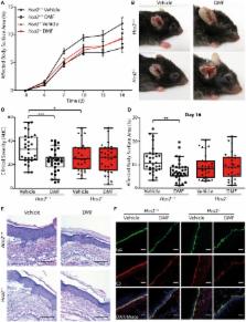
          The drug dimethyl fumarate (DMF) is in clinical use for the treatment of psoriasis and multiple sclerosis. In addition, it has recently been demonstrated to ameliorate skin pathology in mouse models of pemphigoid diseases, a group of autoimmune blistering diseases of the skin and mucous membranes. However, the mode of action of DMF in inflammatory skin diseases has remained elusive. Therefore, we have investigated here the mechanisms by which DMF improves skin pathology, using the antibody transfer model of bullous pemphigoid-like epidermolysis bullosa acquisita (EBA). Experimental EBA was induced by transfer of antibodies against collagen VII that triggered the infiltration of immune cells into the skin and led to inflammatory skin lesions. DMF treatment reduced the infiltration of neutrophils and monocytes into the skin explaining the improved disease outcome in DMF-treated animals. Upon ingestion, DMF is converted to monomethyl fumarate that activates the hydroxycarboxylic acid receptor 2 (HCA 2). Interestingly, neutrophils and monocytes expressed Hca2. To investigate whether the therapeutic effect of DMF in EBA is mediated by HCA 2, we administered oral DMF to Hca2-deficient mice ( Hca2 −/−) and wild-type littermates ( Hca2 +/+) and induced EBA. DMF treatment ameliorated skin lesions in Hca2 +/+ but not in Hca2 −/− animals. These findings demonstrate that HCA 2 is a molecular target of DMF treatment in EBA and suggest that HCA 2 activation limits skin pathology by inhibiting the infiltration of neutrophils and monocytes into the skin.

          Related collections

          Most cited references28

          • Record: found
          • Abstract: found
          • Article: not found

          The β-hydroxybutyrate receptor HCA2 activates a neuroprotective subset of macrophages.

          The ketone body β-hydroxybutyrate (BHB) is an endogenous factor protecting against stroke and neurodegenerative diseases, but its mode of action is unclear. Here we show in a stroke model that the hydroxy-carboxylic acid receptor 2 (HCA2, GPR109A) is required for the neuroprotective effect of BHB and a ketogenic diet, as this effect is lost in Hca2(-/-) mice. We further demonstrate that nicotinic acid, a clinically used HCA2 agonist, reduces infarct size via a HCA2-mediated mechanism, and that noninflammatory Ly-6C(Lo) monocytes and/or macrophages infiltrating the ischemic brain also express HCA2. Using cell ablation and chimeric mice, we demonstrate that HCA2 on monocytes and/or macrophages is required for the protective effect of nicotinic acid. The activation of HCA2 induces a neuroprotective phenotype of monocytes and/or macrophages that depends on PGD2 production by COX1 and the haematopoietic PGD2 synthase. Our data suggest that HCA2 activation by dietary or pharmacological means instructs Ly-6C(Lo) monocytes and/or macrophages to deliver a neuroprotective signal to the brain.
            Bookmark
            • Record: found
            • Abstract: found
            • Article: not found

            PUMA-G and HM74 are receptors for nicotinic acid and mediate its anti-lipolytic effect.

            Nicotinic acid (niacin), a vitamin of the B complex, has been used for almost 50 years as a lipid-lowering drug. The pharmacological effect of nicotinic acid requires doses that are much higher than those provided by a normal diet. Its primary action is to decrease lipolysis in adipose tissue by inhibiting hormone-sensitive triglyceride lipase. This anti-lipolytic effect of nicotinic acid involves the inhibition of cyclic adenosine monophosphate (cAMP) accumulation in adipose tissue through a G(i)-protein-mediated inhibition of adenylyl cyclase. A G-protein-coupled receptor for nicotinic acid has been proposed in adipocytes. Here, we show that the orphan G-protein-coupled receptor, 'protein upregulated in macrophages by interferon-gamma' (mouse PUMA-G, human HM74), is highly expressed in adipose tissue and is a nicotinic acid receptor. Binding of nicotinic acid to PUMA-G or HM74 results in a G(i)-mediated decrease in cAMP levels. In mice lacking PUMA-G, the nicotinic acid-induced decrease in free fatty acid (FFA) and triglyceride plasma levels was abrogated, indicating that PUMA-G mediates the anti-lipolytic and lipid-lowering effects of nicotinic acid in vivo. The identification of the nicotinic acid receptor may be useful in the development of new drugs to treat dyslipidemia.
              Bookmark
              • Record: found
              • Abstract: found
              • Article: not found

              Antigen-Presenting Cells in the Skin.

              Professional antigen-presenting cells (APCs) in the skin include dendritic cells, monocytes, and macrophages. They are highly dynamic, with the capacity to enter skin from the peripheral circulation, patrol within tissue, and migrate through lymphatics to draining lymph nodes. Skin APCs are endowed with antigen-sensing, -processing, and -presenting machinery and play key roles in initiating, modulating, and resolving cutaneous inflammation. Skin APCs are a highly heterogeneous population with functionally specialized subsets that are developmentally imprinted and modulated by local tissue microenvironmental and inflammatory cues. This review explores recent advances that have allowed for a more accurate taxonomy of APC subsets found in both mouse and human skin. It also examines the functional specificity of individual APC subsets and their collaboration with other immune cell types that together promote adaptive T cell and regional cutaneous immune responses during homeostasis, inflammation, and disease.
                Bookmark

                Author and article information

                Contributors
                URI : https://frontiersin.org/people/u/574653
                URI : https://frontiersin.org/people/u/576183
                URI : https://frontiersin.org/people/u/467981
                URI : https://frontiersin.org/people/u/236399
                URI : https://frontiersin.org/people/u/85947
                Journal
                Front Immunol
                Front Immunol
                Front. Immunol.
                Frontiers in Immunology
                Frontiers Media S.A.
                1664-3224
                14 August 2018
                2018
                : 9
                : 1890
                Affiliations
                [1] 1Institute for Experimental and Clinical Pharmacology and Toxicology, University of Lübeck , Lübeck, Germany
                [2] 2Department of Pharmacology, Max Planck Institute for Heart and Lung Research , Bad Nauheim, Germany
                [3] 3Department of Dermatology, Allergy, and Venereology, University of Lübeck , Lübeck, Germany
                Author notes

                Edited by: Angelo Valerio Marzano, Università degli Studi di Milano, Italy

                Reviewed by: Alex Ortega Loayza, Oregon Health & Science University, United States; Jillian M. Richmond, University of Massachusetts Medical School, United States

                *Correspondence: Markus Schwaninger, markus.schwaninger@ 123456pharma.uni-luebeck.de

                These authors have contributed equally to this work.

                Specialty section: This article was submitted to Autoimmune and Autoinflammatory Disorders, a section of the journal Frontiers in Immunology

                Article
                10.3389/fimmu.2018.01890
                6102353
                30154797
                d5a3d11c-f66b-4886-9911-8d7338b936d8
                Copyright © 2018 Wannick, Assmann, Vielhauer, Offermanns, Zillikens, Sadik and Schwaninger.

                This is an open-access article distributed under the terms of the Creative Commons Attribution License (CC BY). The use, distribution or reproduction in other forums is permitted, provided the original author(s) and the copyright owner(s) are credited and that the original publication in this journal is cited, in accordance with accepted academic practice. No use, distribution or reproduction is permitted which does not comply with these terms.

                History
                : 15 June 2018
                : 31 July 2018
                Page count
                Figures: 5, Tables: 1, Equations: 0, References: 33, Pages: 10, Words: 5746
                Funding
                Funded by: Deutsche Forschungsgemeinschaft 10.13039/501100001659
                Award ID: SCHW 416/10-1, SCHW 416/11-1
                Funded by: Exzellenzclusters Entzündungsforschung 10.13039/501100010782
                Categories
                Immunology
                Original Research

                Immunology
                pemphigoid disease,g protein-coupled receptor,immunomodulatory therapy,autoimmune blistering skin disease,neutrophils

                Comments

                Comment on this article