7
views
0
recommends
+1 Recommend
0 collections
    0
    shares
      • Record: found
      • Abstract: found
      • Article: found
      Is Open Access

      Ginsenoside Rg3 inhibits keloid fibroblast proliferation, angiogenesis and collagen synthesis in vitro via the TGF-β/Smad and ERK signaling pathways

      research-article

      Read this article at

      Bookmark
          There is no author summary for this article yet. Authors can add summaries to their articles on ScienceOpen to make them more accessible to a non-specialist audience.

          Abstract

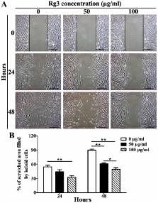
          A wide range of therapeutic options exists for the treatment of keloids, all of which have their own strengths; however, a high risk of side-effects and frequent recurrence remains. Therefore, the present study aimed to identify improved therapeutic approaches or drugs for the treatment of keloids. Ginsenoside Rg3 (Rg3) has been reported to exert numerous antitumor effects, thus indicating that Rg3 may be a potential therapeutic agent that targets keloids. The present study determined the effects of Rg3 on human keloid fibroblasts (KFs) in vitro, and further explored the associated molecular and cellular mechanisms. Keloid scar specimens were obtained from patients, aged between 22 and 35 years, without systemic diseases and primary cells were isolated from keloid tissues. In each assay, KFs were divided into three groups and were cultured in medium with or without various concentrations of Rg3 (50 or 100 μg/ml). Cell viability assay, flow cytometry, quantitative polymerase chain reaction, cell migration assay, immunofluorescence staining, western blot analysis, Transwell cell invasion assay and immunohistochemical analysis were used to analyze the KFs and keloid explant cultures. The results of the present study demonstrated that Rg3 was able to exert an inhibitory effect on the transforming growth factor-β/Smad and extracellular signal-regulated kinase signaling pathways in KFs. The proliferation, migration, invasion, angiogenesis and collagen synthesis of KFs were markedly suppressed following treatment with Rg3. Furthermore, the results of an ex vivo assay indicated that Rg3 inhibited angiogenesis and reduced collagen accumulation in keloids. Significant statistical differences existed between the control and Rg3-treated groups (P<0.05). All of these experimental results suggested that Rg3 may serve as a reliable drug for the treatment of patients with keloids.

          Related collections

          Most cited references41

          • Record: found
          • Abstract: found
          • Article: not found

          Cancer as an overhealing wound: an old hypothesis revisited.

          What is the relationship between the wound-healing process and the development of cancer? Malignant tumours often develop at sites of chronic injury, and tissue injury has an important role in the pathogenesis of malignant disease, with chronic inflammation being the most important risk factor. The development and functional characterization of genetically modified mice that lack or overexpress genes that are involved in repair, combined with gene-expression analysis in wounds and tumours, have highlighted remarkable similarities between wound repair and cancer. However, a few crucial differences were also observed, which could account for the altered metabolism, impaired differentiation capacity and invasive growth of malignant tumours.
            Bookmark
            • Record: found
            • Abstract: found
            • Article: not found

            Identification of Smad7, a TGFbeta-inducible antagonist of TGF-beta signalling.

            TGF-beta signals from the membrane to the nucleus through serine/threonine kinase receptors and their downstream effectors, termed SMAD proteins. The activated TGF-beta receptor induces phosphorylation of two such proteins, Smad2 and Smad3, which form hetero-oligomeric complex(es) with Smad4/DPC4 that translocate to the nucleus, where they then regulate transcriptional responses. However, the mechanisms by which the intracellular signals of TGF-beta are switched off are unclear. Here we report the identification of Smad7, which is related to Smad6. Transfection of Smad7 blocks responses mediated by TGF-beta in mammalian cells, and injection of Smad7 RNA into Xenopus embryos blocks activin/TGF-beta signalling. Smad7 associates stably with the TGF-beta receptor complex, but is not phosphorylated upon TGF-beta stimulation. TGFbeta-mediated phosphorylation of Smad2 and Smad3 is inhibited by Smad7, indicating that the antagonistic effect of Smad7 is exerted at this important regulatory step. TGF-beta rapidly induces expression of Smad7 mRNA, suggesting that Smad7 may participate in a negative feedback loop to control TGF-beta responses.
              Bookmark
              • Record: found
              • Abstract: found
              • Article: not found

              Hyperglycaemic conditions decrease cultured keratinocyte mobility: implications for impaired wound healing in patients with diabetes.

              Elevated blood glucose in patients with diabetes mellitus (DM) leads to complications including poor wound healing. Proper keratinocyte migration and proliferation are the crucial steps during re-epithelialization. We hypothesize that the impaired wound healing in patients with DM is due to the disruption of proper re-epithelialization. We aimed to explore the effects of high glucose on keratinocytes in terms of cell migration and proliferation. Keratinocytes were cultivated in normal and high glucose conditions. Their viability was evaluated by MTS assay. Transwell migration and in vitro scratch assays were used to evaluate their mobility. The mRNA expressions and activities of matrix metalloproteinase (MMP)-2 and MMP-9 were determined. The mRNA of their respective physiological inhibitors, tissue inhibitor of MMP (TIMP)-1 and TIMP-2, was also evaluated. Immunofluorescent staining and Western blotting were used to examine the expression of phosphorylated focal adhesion kinase (pp125(FAK)). The impacts of high glucose on keratinocyte proliferation were assessed by 5-bromo-2'-deoxyuridine incorporation assay. High glucose treatment did not affect keratinocyte viability up to 3 days. In contrast, the mobility of keratinocytes, the activities and gene expressions of MMP-2 and MMP-9, the expression of pp125(FAK), and the cell proliferation after 5 days were significantly downregulated after hyperglycaemic treatments while the mRNA expression of TIMP-1 increased. Under hyperglycaemic conditions, keratinocytes demonstrate reduced migration and decreased proliferation capacities. These impairments of keratinocyte functions are likely to result in inadequate re-epithelialization. These defective physiological events provide a reasonable explanation for the poor wound healing commonly observed in patients with DM.
                Bookmark

                Author and article information

                Journal
                Int J Mol Med
                Int. J. Mol. Med
                IJMM
                International Journal of Molecular Medicine
                D.A. Spandidos
                1107-3756
                1791-244X
                March 2018
                04 January 2018
                04 January 2018
                : 41
                : 3
                : 1487-1499
                Affiliations
                Department of Plastic and Reconstructive Surgery, Shanghai Ninth People's Hospital Affiliated Shanghai Jiao Tong University School of Medicine, Shanghai 200011, P.R. China
                Author notes
                Correspondence to: Dr Yuguang Zhang or Dr Wenbo Wang, Department of Plastic and Reconstructive Surgery, Shanghai Ninth People's Hospital Affiliated Shanghai Jiao Tong University School of Medicine, 639 Zhizaoju Road, Shanghai 200011, P.R. China, E-mail: zhangyg18@ 123456126.com , E-mail: wangwenbo0903@ 123456126.com
                [*]

                Contributed equally

                Article
                ijmm-41-03-1487
                10.3892/ijmm.2018.3362
                5819908
                29328420
                d6baddae-3b8f-4589-89ed-1c256683d070
                Copyright: © Tang et al.

                This is an open access article distributed under the terms of the Creative Commons Attribution-NonCommercial-NoDerivs License, which permits use and distribution in any medium, provided the original work is properly cited, the use is non-commercial and no modifications or adaptations are made.

                History
                : 15 November 2016
                : 15 December 2017
                Categories
                Articles

                wound healing,keloid,ginsenoside rg3,signaling pathways
                wound healing, keloid, ginsenoside rg3, signaling pathways

                Comments

                Comment on this article