6
views
0
recommends
+1 Recommend
0 collections
    0
    shares
      • Record: found
      • Abstract: found
      • Article: not found

      T 1- and T 2-weighted Magnetic Resonance Dual Contrast by Single Core Truncated Cubic Iron Oxide Nanoparticles with Abrupt Cellular Internalization and Immune Evasion

      research-article

      Read this article at

      ScienceOpenPublisherPMC
      Bookmark
          There is no author summary for this article yet. Authors can add summaries to their articles on ScienceOpen to make them more accessible to a non-specialist audience.

          Abstract

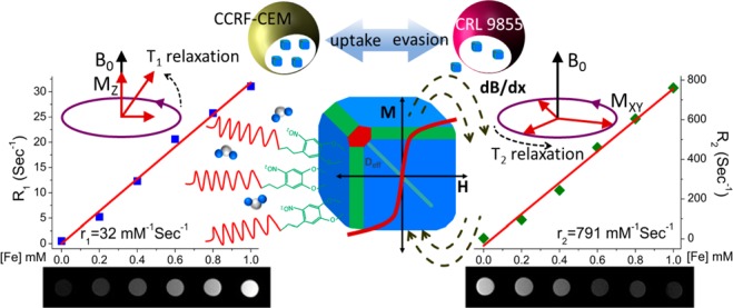
          Conventional T 1- or T 2-weighted single mode contrast-enhanced magnetic resonance imaging (MRI) may produce false results. Thereby, there is a need to develop dual contrast agents, T 1- and T 2-weighted, for more accurate MRI imaging. The dual contrast agents should possess high magnetic resonance (MR) relaxivities, targeted tumor linking, and minimum recognition by the immune system. We have developed nitrodopamine-PEG grafted single core truncated cubic iron oxide nanoparticles (ND-PEG-tNCIOs) capable of producing marked dual contrasts in MRI with enhanced longitudinal and transverse relaxivities of 32 ± 1.29 and 791 ± 38.39 mM –1 s –1, respectively. Furthermore, the ND-PEG-tNCIOs show excellent colloidal stability in physiological buffers and higher cellular internalization in cancerous cells than in phagocytic cells, indicating the immune evasive capability of the nanoparticles. These findings indicate that tNCIOs are strong candidates for dual contrast MRI imaging, which is vital for noninvasive real-time detection of nascent cancer cells in vivo and for monitoring stem cells transplants.

          Related collections

          Most cited references53

          • Record: found
          • Abstract: found
          • Article: not found

          Regulated portals of entry into the cell.

          The plasma membrane is the interface between cells and their harsh environment. Uptake of nutrients and all communication among cells and between cells and their environment occurs through this interface. 'Endocytosis' encompasses several diverse mechanisms by which cells internalize macromolecules and particles into transport vesicles derived from the plasma membrane. It controls entry into the cell and has a crucial role in development, the immune response, neurotransmission, intercellular communication, signal transduction, and cellular and organismal homeostasis. As the complexity of molecular interactions governing endocytosis are revealed, it has become increasingly clear that it is tightly coordinated and coupled with overall cell physiology and thus, must be viewed in a broader context than simple vesicular trafficking.
            Bookmark
            • Record: found
            • Abstract: found
            • Article: not found

            Role of target geometry in phagocytosis.

            Phagocytosis is a principal component of the body's innate immunity in which macrophages internalize targets in an actin-dependent manner. Targets vary widely in shape and size and include particles such as pathogens and senescent cells. Despite considerable progress in understanding this complicated process, the role of target geometry in phagocytosis has remained elusive. Previous studies on phagocytosis have been performed using spherical targets, thereby overlooking the role of particle shape. Using polystyrene particles of various sizes and shapes, we studied phagocytosis by alveolar macrophages. We report a surprising finding that particle shape, not size, plays a dominant role in phagocytosis. All shapes were capable of initiating phagocytosis in at least one orientation. However, the local particle shape, measured by tangent angles, at the point of initial contact dictates whether macrophages initiate phagocytosis or simply spread on particles. The local shape determines the complexity of the actin structure that must be created to initiate phagocytosis and allow the membrane to move over the particle. Failure to create the required actin structure results in simple spreading and not internalization. Particle size primarily impacts the completion of phagocytosis in cases where particle volume exceeds the cell volume.
              Bookmark
              • Record: found
              • Abstract: found
              • Article: not found

              Superparamagnetic iron oxide: pharmacokinetics and toxicity.

              The pharmacokinetics (distribution, metabolism, bioavailability, excretion) and toxicity (acute and subacute toxicity, mutagenicity) of a superparamagnetic iron oxide preparation (AMI-25), currently used in clinical trials, were evaluated by 59Fe radiotracer studies, measurements of relaxation times, the ability to reverse iron deficiency anemia, histologic examination, and laboratory parameters. One hour after administration of AMI-25 to rats (18 mumol Fe/kg; 1 mg Fe/kg), 82.6 +/- 0.3% of the administered dose was sequestered in the liver and 6.2 +/- 7.6% in the spleen. Peak concentrations of 59Fe were found in liver after 2 hr and in the spleen after 4 hr. 59Fe slowly cleared from liver (half-life, 3 days) and spleen (half-life, 4 days) and was incorporated into hemoglobin of erythrocytes in a time-dependent fashion. The half-time of the T2 effect on liver and spleen (24-48 hr) was shorter than the 59Fe clearance, indicating metabolism of AMI-25 into other forms of iron. IV administration of AMI-25 (30 mg Fe/kg) corrected iron-deficiency anemia and showed bioavailability similar to that of commercially available IV iron preparations within 7 days. No acute or subacute toxic effects were detected by histologic or serologic studies in rats or beagle dogs who received a total of 3000 mumol Fe/kg, 150 times the dose proposed for MR imaging of the liver. Our results indicate that AMI-25 is a fully biocompatible potential contrast agent for MR.
                Bookmark

                Author and article information

                Journal
                ACS Appl Bio Mater
                ACS Appl Bio Mater
                mt
                aabmcb
                ACS Applied Bio Materials
                American Chemical Society
                2576-6422
                15 June 2018
                16 July 2018
                : 1
                : 1
                : 79-89
                Affiliations
                []Molecular Sciences Research Center, University of Puerto Rico , San Juan, Puerto Rico 00926-2614, United States
                []Department of Physics, University of Puerto Rico, Río Piedras Campus , San Juan, Puerto Rico 00925-2537, United States
                []Department of Chemistry, University of Puerto Rico, Río Piedras Campus , San Juan, Puerto Rico 00925-2537, United States
                []Department of Biology, University of Puerto Rico , Río Piedras Campus, San Juan, Puerto Rico 00925-2537, United States
                [§ ]Department of Environmental Health, Harvard University , Boston, Massachusetts 02115-5810, United States
                []School of Electrical Engineering and Computer Science, Ohio University , Athens, Ohio 45701-2769, United States
                Author notes
                Article
                10.1021/acsabm.8b00016
                6077774
                d704be05-da62-411c-a22d-83d4e2093b23
                Copyright © 2018 American Chemical Society

                This is an open access article published under an ACS AuthorChoice License, which permits copying and redistribution of the article or any adaptations for non-commercial purposes.

                History
                : 21 April 2018
                : 15 June 2018
                Categories
                Article
                Custom metadata
                mt8b00016
                mt-2018-00016z

                magnetic resonance imaging,t1- and t2-weighted dual contrast,longitudinal and transverse relaxivity,truncated cubic iron oxide nanoparticles,cellular internalization,immune evasion

                Comments

                Comment on this article