9
views
0
recommends
+1 Recommend
0 collections
    0
    shares
      • Record: found
      • Abstract: found
      • Article: found
      Is Open Access

      Trans-Sellar Trans-Sphenoidal Herniation of Third Ventricle with Cleft Palate and Microophthalmia: Report of a Case and Review of Literature

      case-report

      Read this article at

      Bookmark
          There is no author summary for this article yet. Authors can add summaries to their articles on ScienceOpen to make them more accessible to a non-specialist audience.

          Abstract

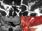
          Trans -sellar trans-sphenoidal encephalocele is an extremely rare entity. We present the case of an 18-month old boy who presented with a trans-sellar, trans-sphenoidal encephalocele associated with cleft lip, cleft palate and microphthalmia. This patient was treated successfully by a trans-cranial extra-dural route. In this paper, we discuss the clinico-radiological findings as well as various surgical options in managing these rare lesions and briefly review the literature.

          Related collections

          Most cited references21

          • Record: found
          • Abstract: found
          • Article: found
          Is Open Access

          Sternberg's canal as a cause of encephalocele within the lateral recess of the sphenoid sinus: A report of two cases

          Background: Intrasphenoidal encephaloceles are extremely rare findings. Sternberg's canal is a lateral craniopharyngeal canal resulting from incomplete fusion of the greater wings of the sphenoid bone with the basisphenoid. It acts as a weak spot of the skull base, which may lead to develop a temporal lobe encephalocele protruding into the lateral recess of the sphenoid sinus (SS). Case Description: We present two cases of intrasphenoidal encephalocele due to persistence of the lateral craniopharyngeal canal. The first case presented with cerebrospinal fluid (CSF) rhinorrhea and the second one was referred to the neurosurgical department with CSF rhinorrhea and meningitis. Radiological investigations consisted of computed tomography (CT) scan, CT cisternography and magnetic resonance images in both cases. These imaging studies identified a herniated temporal lobe through a bony defect which communicates the middle cranial fossa with the lateral recess of the SS. Both patients underwent a transcranial repair of the encephalocele because of the previous failure of the endoscopic surgery. There was no complication related to the surgical procedure and no recurrence of CSF leakage occurred 2 and 3 years after surgery, respectively. Conclusion: Encephalocele within the lateral recess of the SS is a rare entity which must be suspected in patients who present with spontaneous CSF rhinorrhea. Congenital intrasphenoidal encephaloceles, which are located medial to the foramen rotundum, seem to be due to persistence of the Sternberg's canal. Transcranial approach is a good option when a transnasal approach had failed previously.
            Bookmark
            • Record: found
            • Abstract: found
            • Article: not found

            Intrasphenoidal encephaloceles--a clinical entity.

            In the current classification basal encephaloceles are grouped together with real transsphenoidal encephaloceles. But those encephaloceles extending only into but not through the sphenoid sinus seem to represent a specific clinical entity and therefore should be regarded as a rare subgroup of sphenoidal encephaloceles. One personal case and six cases from the literature are reviewed, the own case being associated with an empty sella turcica. The initial sign is rhinorrhea, almost invariably. The association with other intracranial anomalies is uncommon. The extradural transsphenoidal or transethmoidal midline approach accompanied by a shunting procedure today is the most suitable method of surgical treatment.
              Bookmark
              • Record: found
              • Abstract: found
              • Article: not found

              Transsphenoidal meningoencephalocele.

              Encephaloceles are rare cranio-facial developmental anomalies. In this paper, we discuss the case of an 8-month-old girl affected by a transsphenoidal meningoencephalocele and try to give landmarks of the state of art in the surgical treatment of this rare pathology.
                Bookmark

                Author and article information

                Journal
                Asian J Neurosurg
                Asian J Neurosurg
                AJNS
                Asian Journal of Neurosurgery
                Medknow Publications & Media Pvt Ltd (India )
                1793-5482
                2248-9614
                Jul-Sep 2018
                : 13
                : 3
                : 782-785
                Affiliations
                [1] Department of Neurosurgery, SGPGIMS, Lucknow, Uttar Pradesh, India
                Author notes
                Address for correspondence: Dr. Sanjay Behari, Department of Neurosurgery, SGPGIMS, Raibareilly Road, Lucknow - 226 014, Uttar Pradesh, India. E-mail: sbehari27@ 123456yahoo.com
                Article
                AJNS-13-782
                10.4103/1793-5482.238003
                6159010
                30283546
                d7522cc0-4251-44ba-981b-9e0947ccba1c
                Copyright: © 2018 Asian Journal of Neurosurgery

                This is an open access journal, and articles are distributed under the terms of the Creative Commons Attribution-NonCommercial-ShareAlike 4.0 License, which allows others to remix, tweak, and build upon the work non-commercially, as long as appropriate credit is given and the new creations are licensed under the identical terms.

                History
                Categories
                Case Report

                Surgery
                basal encephalocele,cleft lip,craniopharyngeal canal,transcranial surgery,trans-sellar,trans-sphenoidal

                Comments

                Comment on this article