18
views
0
recommends
+1 Recommend
0 collections
    0
    shares
      • Record: found
      • Abstract: found
      • Article: found
      Is Open Access

      Pseudo cardiac tamponade in the setting of excess pericardial fat

      research-article

      Read this article at

      Bookmark
          There is no author summary for this article yet. Authors can add summaries to their articles on ScienceOpen to make them more accessible to a non-specialist audience.

          Abstract

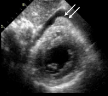
          Cardiac tamponade is the phenomenon of hemodynamic compromise caused by a pericardial effusion. Following a myocardial infarction, the most common causes of pericardial fluid include early pericarditis, Dressler's syndrome, and hemopericardium secondary to a free wall rupture. On transthoracic echocardiography, pericardial fluid appears as an echo-free space in between the visceral and parietal layers of the pericardium. Pericardial fat has a similar appearance on echocardiography and it may be difficult to discern the two entities. We present a case of a post-MI patient demonstrating pseudo tamponade physiology in the setting of excessive pericardial fat.

          Related collections

          Most cited references18

          • Record: found
          • Abstract: found
          • Article: not found

          Pericardial fat, visceral abdominal fat, cardiovascular disease risk factors, and vascular calcification in a community-based sample: the Framingham Heart Study.

          Pericardial fat may be an important mediator of metabolic risk. Correlations with cardiovascular disease risk factors and vascular calcification in a community-based sample are lacking. We sought to examine associations between pericardial fat, metabolic risk factors, and vascular calcification. Participants free of cardiovascular disease from the Framingham Heart Study (n=1155, mean age 63 years, 54.8% women) who were part of a multidetector computed tomography study underwent quantification of intrathoracic fat, pericardial fat, visceral abdominal fat (VAT), coronary artery calcification, and aortic artery calcification. Intrathoracic and pericardial fat volumes were examined in relation to body mass index, waist circumference, VAT, metabolic risk factors, coronary artery calcification, and abdominal aortic calcification. Intrathoracic and pericardial fat were directly correlated with body mass index (r=0.41 to 0.51, P 0.05). Pericardial fat, but not intrathoracic fat, was associated with coronary artery calcification after multivariable and VAT adjustment (odds ratio 1.21, 95% confidence interval 1.005 to 1.46, P=0.04), whereas intrathoracic fat, but not pericardial fat, was associated with abdominal aortic calcification (odds ratio 1.32, 95% confidence interval 1.03 to 1.67, P=0.03). Pericardial fat is correlated with multiple measures of adiposity and cardiovascular disease risk factors, but VAT is a stronger correlate of most metabolic risk factors. However, intrathoracic and pericardial fat are associated with vascular calcification, which suggests that these fat depots may exert local toxic effects on the vasculature.
            Bookmark
            • Record: found
            • Abstract: found
            • Article: not found

            Human epicardial adipose tissue: a review.

            We discuss the anatomy, physiology, and pathophysiology of epicardial adipose tissue and its relationship to coronary atherosclerosis. Epicardial fat stores triglyceride to supply free fatty acids for myocardial energy production and produces adipokines. It shares a common embryological origin with mesenteric and omental fat. Like visceral abdominal fat, epicardial fat thickness, measured by echocardiography, is increased in obesity. Epicardial fat could influence coronary atherogenesis and myocardial function because there is no fibrous fascial layer to impede diffusion of free fatty acids and adipokines between it and the underlying vessel wall as well as the myocardium. Segments of coronary arteries lacking epicardial fat or separated from it by a bridge of myocardial tissue are protected against the development of atherosclerosis in those segments. However, when epicardial fat is totally absent in congenital generalized lipodystrophy, coronary atherosclerosis can still occur. Macrophages are more numerous and densely packed in the periadventitial fat of human atherosclerotic coronary arteries with lipid cores than in that of fibrocalcific or nonatherosclerotic coronary arteries. In obese patients with multiple cardiovascular risk factors, epicardial fat around atheromatous coronaries secretes several proinflammatory cytokines and is infiltrated by macrophages, lymphocytes, and basophils. Epicardial adipokine expression in obesity without coronary atherosclerosis has not been determined. In nonobese patients, epicardial fat around atheromatous coronary arteries expresses proinflammatory cytokines but produces either less adiponectin, a vasoprotective adipokine, than fat around nonatheromatous coronaries or a similar amount compared with thoracic subcutaneous fat. Further studies should be done to test the hypothesis that adipokines produced by and released from human epicardial adipose tissue might contribute locally to the pathogenesis of coronary atherosclerosis.
              Bookmark
              • Record: found
              • Abstract: found
              • Article: not found

              Relationship of epicardial adipose tissue by echocardiography to coronary artery disease.

              To study the relationship of echocardiographic epicardial adipose tissue (EAT) with coronary artery disease (CAD) risk factors and the extent of coronary atherosclerosis. EAT thickness was measured in 527 patients undergoing their first coronary angiography. EAT was defined as an echo-lucent area on the free wall of the right ventricle on the still image of the two-dimensional echocardiogram at end diastole in the parasternal long-axis and parasternal short-axis views. A CT scan at the umbilicus was acquired to measure abdominal visceral adipose tissue (VAT) from a random sample of 30 patients. The extent of coronary atherosclerosis was assessed using a coronary atherosclerosis score based on the quantitative coronary angiography results. EAT thickness was correlated with abdominal VAT (r(s) = 0.626, p or =3.0 mm) was an independent factor of CAD on multiple logistic analysis (odds ratio = 3.357; 95% CI 2.177 to 5.175, p<0.001). These results suggest that EAT may reflect the amount of visceral fat, which is associated with insulin resistance and inflammation. The echocardiographic measurement of EAT may provide additional information for assessing CAD risk and predicting the extent and activity of CAD.
                Bookmark

                Author and article information

                Journal
                Cardiovasc Ultrasound
                Cardiovascular Ultrasound
                BioMed Central
                1476-7120
                2009
                22 January 2009
                : 7
                : 3
                Affiliations
                [1 ]Section of Cardiology, Department of Cardiac Sciences, University of Manitoba, Winnipeg, Manitoba, Canada
                [2 ]Section of Cardiac Surgery, Department of Cardiac Sciences, University of Manitoba, Winnipeg, Manitoba, Canada
                [3 ]Institute of Cardiovascular Sciences, St. Boniface Research Centre, University of Manitoba, Winnipeg, Manitoba, Canada
                [4 ]Department of Radiology, University of Manitoba, Winnipeg, Manitoba, Canada
                Article
                1476-7120-7-3
                10.1186/1476-7120-7-3
                2632988
                19161596
                d899328d-1a34-48f6-8fb0-ff760cc64f2b
                Copyright © 2009 Nguyen et al; licensee BioMed Central Ltd.

                This is an Open Access article distributed under the terms of the Creative Commons Attribution License ( http://creativecommons.org/licenses/by/2.0), which permits unrestricted use, distribution, and reproduction in any medium, provided the original work is properly cited.

                History
                : 5 January 2009
                : 22 January 2009
                Categories
                Case Report

                Cardiovascular Medicine
                Cardiovascular Medicine

                Comments

                Comment on this article