5
views
0
recommends
+1 Recommend
0 collections
    0
    shares
      • Record: found
      • Abstract: found
      • Article: found
      Is Open Access

      Cardiac Tumors : JACC CardioOncology State-of-the-Art Review

      review-article
      , MBBS, BSc a , , BMed a , b , , MBChB, MD a , , MD c , , MD d , , PhD a , , MD a , , MBBS, MSc, PhD a , b , , , MD e , f , ,
      JACC: CardioOncology
      Elsevier
      cardiac masses, cardiac metastases, cardiac myxoma, cardiac sarcoma, cardiac tumors, papillary fibroelastoma, pericardial cyst, pericardial mesothelioma, pericardial tumors, CMR, cardiovascular magnetic resonance imaging, CT, computed tomography, FDG, fluorodeoxyglucose, PCT, primary cardiac tumor, PET, positron emission tomography, RT3DE, real-time 3-dimensional echocardiography, TEE, transesophageal echocardiography, TTE, transthoracic echocardiography, UEA, ultrasound-enhancing agent

      Read this article at

      Bookmark
          There is no author summary for this article yet. Authors can add summaries to their articles on ScienceOpen to make them more accessible to a non-specialist audience.

          Abstract

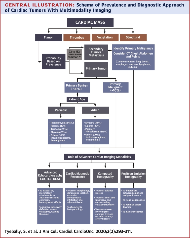
          Cardiac masses are rare, but remain an important component of cardio-oncology practice. These include benign tumors, malignant tumors (primary and secondary) and tumor-like conditions (e.g., thrombus, Lambl’s excrescences, and pericardial cyst). The advent of multimodality imaging has enabled identification of the etiology of cardiac masses in many cases, especially in conjunction with information from clinical settings. This paper provides a comprehensive review of the epidemiology, clinical presentation, imaging, diagnosis, management, and outcomes of cardiac masses.

          Central Illustration

          Highlights

          • Cardiac tumors are rare and should be considered as part of the differential diagnosis of any space-occupying mass noted on cardiovascular and/or thoracic imaging modalities.

          • It may be possible to get close to a diagnosis without biopsy using a structured imaging approach.

          • The prognosis and treatment of each tumor is different, although early diagnosis is usually associated with a better outcome.

          Related collections

          Most cited references122

          • Record: found
          • Abstract: found
          • Article: not found

          Cardiac tumours: diagnosis and management.

          Primary cardiac tumours are rare, with an autopsy incidence ranging from 0.001% to 0.030%. Three-quarters of these tumours are benign and nearly half of the benign tumours are myxomas. Metastases to the heart are far more common than primary cardiac tumours. Primary cardiac tumours present with one or more of the symptoms of the classic triad of: cardiac symptoms and signs resulting from intracardiac obstruction; signs of systemic embolisation; and systemic or constitutional symptoms. They are diagnosed by use of transthoracic and transoesophageal echocardiograms, MRI, and CT scan. Whereas surgery is indicated in patients with benign tumours, systemic chemotherapy is indicated in those who have widespread or unresectable malignant disease, and chemotherapy and radiotherapy are usually combined in treatment of patients with primary cardiac lymphomas. The prognosis after surgery is usually excellent in the case of benign tumours but is unfortunately still limited in localised malignant diseases. Patients with sarcomas live for a mean of 3 months to 1 year, and those with lymphomas live up to 5 years if treated, but usually die within 1 month if untreated.
            Bookmark
            • Record: found
            • Abstract: found
            • Article: not found

            Clinical presentation of left atrial cardiac myxoma. A series of 112 consecutive cases.

            We analyzed a series of 112 consecutive cases of left atrial myxoma diagnosed in a single French hospital (72 women and 40 men; age range, 5-84 yr) over 40 years, from 1959 to 1998. Symptoms of mitral valve obstruction, the first arm of the classic triad of myxoma presentation, were present in 75 patients (67%), with mostly cardiac failure or malaise. Symptoms of embolism, the second frequent presentation in the classic triad, were observed in 33 cases (29%) with 1 or several locations, essentially cerebral emboli with stroke. Males are statistically at greater risk than females of developing embolic complications. The third arm of the classic triad consists of constitutional symptoms (34%) with fever, weight loss, or symptoms resembling connective tissue disease, due to cytokine (interleukin-6) secretion. Younger and male patients have more neurologic symptoms, and female patients have more systemic symptoms. Seventy-two patients (64%) had cardiac auscultation abnormalities, essentially pseudo-mitral valve disease (53.5%) and more rarely the suggestive tumor plop (15%). The most frequent electrocardiographic sign was left atrial hypertrophy (35%), whereas arrhythmias were uncommon. The greater number of myxoma patients (98) diagnosed preoperatively after 1977 reflects the introduction of echocardiography as a noninvasive diagnostic procedure. However, there was no significant reduction in the average time from onset of symptoms to operation between patients seen in the periods before and after 1977. The tumor diameter ranged from 1 to 15 cm with a weight of between 15 and 180 g (mean, 37 g). The myxoma surface was friable or villous in 35% of the cases, and smooth in the other 65% cases. Myxomas in patients presenting with embolism have a friable surface; those in patients with cardiac symptoms, pseudo-mitral auscultation signs, tumor plop, and electrocardiogram or radiologic signs of left atrium hypertrophy and dilatation are significantly the larger tumors. The long-term prognosis is excellent, and only 4 deaths occurred among our 112 cases over a median follow-up of 3 years. The recurrence rate is low (5%), but long-term follow-up and serial echocardiography are advisable especially for young patients.
              Bookmark
              • Record: found
              • Abstract: found
              • Article: not found

              Tumors of the heart. A 20-year experience with a review of 12,485 consecutive autopsies.

              Cardiac involvement by primary and secondary tumors is one of the least investigated subjects in oncology. Seven cases of primary and 154 cases of secondary cardiac tumors from autopsies performed over a 20-year period (1972 through 1991) at Queen Mary Hospital, Hong Kong, were reviewed. During this period, 12,485 autopsies were performed, and the autopsy incidence for primary and secondary heart tumors is thus 0.056% and 1.23%, respectively. Only seven primary cardiac tumors were found, including two myxomas, two rhabdomyomas, two hemangiomas, and one lipoma. For secondary tumors involving the heart (including both metastasis and local extension), important primary tumors in male subjects were carcinoma of the lung (31.7%), esophageal carcinoma (28.7%), lymphoma (11.9%), carcinoma of the liver (6.9%), leukemia (4.0%), and gastric carcinoma (4.0%), while in female subjects, carcinoma of the lung (35.9%), lymphoma (17.0%), carcinoma of the breast (7.5%), and pancreatic carcinoma (7.5%) predominated. Overall, the three most common malignant neoplasms encountered were carcinoma of the lung, esophageal carcinoma, and lymphoma. Pericardium, including epicardium, was the most common location of cardiac involvement by secondary tumors, followed by myocardium and endocardium. The present study showed a higher percentage of esophageal carcinoma and carcinoma of the liver (reflecting the higher incidence of these tumors in Hong Kong Chinese), but a lower incidence of carcinoma of the breast when compared with other series. The metastatic lung tumors showed an unusual predominance of adenocarcinoma.
                Bookmark

                Author and article information

                Contributors
                @arjunkg
                @avirupguha
                Journal
                JACC CardioOncol
                JACC CardioOncol
                JACC: CardioOncology
                Elsevier
                2666-0873
                16 June 2020
                June 2020
                16 June 2020
                : 2
                : 2
                : 293-311
                Affiliations
                [a ]Cardiology Department, Barts Heart Centre, St Bartholomew’s Hospital, London, United Kingdom
                [b ]Cardio-Oncology Service, University College London Hospital, London, United Kingdom
                [c ]Jordan University of Science and Technology, Al Ramtha, Irbid, Jordan
                [d ]Division of Cardiology, Loyola University Medical Center, Chicago, Illinois, USA
                [e ]Cardio-Oncology Program, Division of Cardiology, Ohio State University, Columbus, Ohio, USA
                [f ]Harrington Heart and Vascular Institute, Case Western Reserve University, Cleveland, Ohio, USA
                Author notes
                [] Address for correspondence: Dr. Avirup Guha, Harrington Heart and Vascular Institute, Case Western Reserve University, 350 Hillcrest Drive, Upper Level, Ashland, Ohio 44805. avirup.guha@ 123456uhhospitals.org @avirupguha
                [∗]

                Drs. Ghosh and Guha contributed equally to the manuscript.

                Article
                S2666-0873(20)30091-0
                10.1016/j.jaccao.2020.05.009
                8352246
                34396236
                da959344-f624-4084-b8b8-6c6c15ef3dc1
                © 2020 The Authors

                This is an open access article under the CC BY-NC-ND license (http://creativecommons.org/licenses/by-nc-nd/4.0/).

                History
                : 28 April 2020
                : 7 May 2020
                : 11 May 2020
                Categories
                State-of-the-Art Review

                cardiac masses,cardiac metastases,cardiac myxoma,cardiac sarcoma,cardiac tumors,papillary fibroelastoma,pericardial cyst,pericardial mesothelioma,pericardial tumors,cmr, cardiovascular magnetic resonance imaging,ct, computed tomography,fdg, fluorodeoxyglucose,pct, primary cardiac tumor,pet, positron emission tomography,rt3de, real-time 3-dimensional echocardiography,tee, transesophageal echocardiography,tte, transthoracic echocardiography,uea, ultrasound-enhancing agent

                Comments

                Comment on this article