55
views
0
recommends
+1 Recommend
0 collections
    0
    shares
      • Record: found
      • Abstract: found
      • Article: found
      Is Open Access

      Developmental timing of CCM2 loss influences cerebral cavernous malformations in mice

      research-article

      Read this article at

      Bookmark
          There is no author summary for this article yet. Authors can add summaries to their articles on ScienceOpen to make them more accessible to a non-specialist audience.

          Abstract

          As revealed in a new model of cerebral cavernous malformations (CCM), the timing of ablation of Ccm genes determines whether or not CCM lesions arise in brain and retina venous beds.

          Abstract

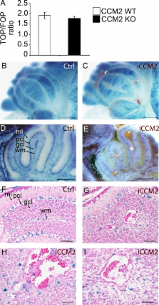
          Cerebral cavernous malformations (CCM) are vascular malformations of the central nervous system (CNS) that lead to cerebral hemorrhages. Familial CCM occurs as an autosomal dominant condition caused by loss-of-function mutations in one of the three CCM genes. Constitutive or tissue-specific ablation of any of the Ccm genes in mice previously established the crucial role of Ccm gene expression in endothelial cells for proper angiogenesis. However, embryonic lethality precluded the development of relevant CCM mouse models. Here, we show that endothelial-specific Ccm2 deletion at postnatal day 1 (P1) in mice results in vascular lesions mimicking human CCM lesions. Consistent with CCM1/3 involvement in the same human disease, deletion of Ccm1/3 at P1 in mice results in similar CCM lesions. The lesions are located in the cerebellum and the retina, two organs undergoing intense postnatal angiogenesis. Despite a pan-endothelial Ccm2 deletion, CCM lesions are restricted to the venous bed. Notably, the consequences of Ccm2 loss depend on the developmental timing of Ccm2 ablation. This work provides a highly penetrant and relevant CCM mouse model.

          Related collections

          Most cited references45

          • Record: found
          • Abstract: found
          • Article: not found

          Molecular distinction and angiogenic interaction between embryonic arteries and veins revealed by ephrin-B2 and its receptor Eph-B4.

          The vertebrate circulatory system is composed of arteries and veins. The functional and pathological differences between these vessels have been assumed to reflect physiological differences such as oxygenation and blood pressure. Here we show that ephrin-B2, an Eph family transmembrane ligand, marks arterial but not venous endothelial cells from the onset of angiogenesis. Conversely, Eph-B4, a receptor for ephrin-B2, marks veins but not arteries. ephrin-B2 knockout mice display defects in angiogenesis by both arteries and veins in the capillary networks of the head and yolk sac as well as in myocardial trabeculation. These results provide evidence that differences between arteries and veins are in part genetically determined and suggest that reciprocal signaling between these two types of vessels is crucial for morphogenesis of the capillary beds.
            Bookmark
            • Record: found
            • Abstract: found
            • Article: not found

            Mapping Wnt/beta-catenin signaling during mouse development and in colorectal tumors.

            Wntbeta-catenin signaling plays key roles in several developmental and pathological processes. Domains of Wnt expression have been extensively investigated in the mouse, but the tissues receiving the signal remain largely unidentified. To define which cells respond to activated beta-catenin during mammalian development, we generated the beta-catenin-activated transgene driving expression of nuclear beta-galactosidase reporter (BAT-gal) transgenic mice, expressing the lacZ gene under the control of beta-cateninT cell factor responsive elements. Reporter gene activity is found in known organizing centers, such as the midhindbrain border and the limb apical ectodermal ridge. Moreover, BAT-gal expression identifies novel sites of Wnt signaling, like notochord, endothelia, and areas of the adult brain, revealing an unsuspected dynamic pattern of beta-catenin transcriptional activity. Expression of the transgene was analyzed in mutant backgrounds. In lipoprotein receptor-related protein 6-null homozygous mice, which lack a Wnt coreceptor, BAT-gal staining is absent in mutant tissues, indicating that BAT-gal mice are bona fide in vivo indicators of Wntbeta-catenin signaling. Analyses of BAT-gal expression in the adenomatous polyposis coli (multiple intestinal neoplasia+) background revealed betacatenin transcriptional activity in intestinal adenomas but surprisingly not in normal crypt cells. In summary, BAT-gal mice unveil the entire complexity of Wntbeta-catenin signaling in mammals and have broad application potentials for the identification of Wnt-responsive cell populations in development and disease.
              Bookmark
              • Record: found
              • Abstract: found
              • Article: not found

              Endothelial adherens junctions control tight junctions by VE-cadherin-mediated upregulation of claudin-5.

              Intercellular junctions mediate adhesion and communication between adjoining cells. Although formed by different molecules, tight junctions (TJs) and adherens junctions (AJs) are functionally and structurally linked, but the signalling pathways behind this interaction are unknown. Here we describe a cell-specific mechanism of crosstalk between these two types of structure. We show that endothelial VE-cadherin at AJs upregulates the gene encoding the TJ adhesive protein claudin-5. This effect requires the release of the inhibitory activity of forkhead box factor FoxO1 and the Tcf-4-beta-catenin transcriptional repressor complex. Vascular endothelial (VE)-cadherin acts by inducing the phosphorylation of FoxO1 through Akt activation and by limiting the translocation of beta-catenin to the nucleus. These results offer a molecular basis for the link between AJs and TJs and explain why VE-cadherin inhibition may cause a marked increase in permeability.
                Bookmark

                Author and article information

                Journal
                J Exp Med
                J. Exp. Med
                jem
                The Journal of Experimental Medicine
                The Rockefeller University Press
                0022-1007
                1540-9538
                29 August 2011
                : 208
                : 9
                : 1835-1847
                Affiliations
                [1 ]Institut National de la Santé et de la Recherche Médicale, UMR-S 740, 75010 Paris, France
                [2 ]Université Paris 7-Denis Diderot, Faculté de Médecine, Site Lariboisière, Paris, F-75010, France
                [3 ]Fondazione Italiana per la Ricerca sul Cancro Institute of Molecular Oncology (IFOM), 20139 Milano, Italy
                [4 ]AP-HP, Groupe Hospitalier Saint-Louis Lariboisiere-Fernand-Widal, Paris, F-75010, France
                [5 ]Department of Pathology, Centre Hospitalier Universitaire, Avenue Côte de Nacre, 14032 Caen, France
                [6 ]Max Planck Institute for Molecular Biomedicine, Faculty of Medicine, Department of Tissue Morphogenesis and University of Münster, 48149 Münster, Germany
                [7 ]Department of Biomolecular Sciences and Biotechnologies, Milan University School of Sciences, 20133 Milan, Italy
                Author notes
                CORRESPONDENCE Elisabeth Tournier-Lasserve: tournier-lasserve@ 123456univ-paris-diderot.fr

                N. Rudini and L. Maddaluno contributed equally to this paper.

                Article
                20110571
                10.1084/jem.20110571
                3171098
                21859843
                db11c5e3-286a-435d-8bb6-118ae9384c50
                © 2011 Boulday et al.

                This article is distributed under the terms of an Attribution–Noncommercial–Share Alike–No Mirror Sites license for the first six months after the publication date (see http://www.rupress.org/terms). After six months it is available under a Creative Commons License (Attribution–Noncommercial–Share Alike 3.0 Unported license, as described at http://creativecommons.org/licenses/by-nc-sa/3.0/).

                History
                : 23 March 2011
                : 26 July 2011
                Categories
                Article

                Medicine
                Medicine

                Comments

                Comment on this article