- Record: found
- Abstract: found
- Article: found
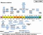
A Combination of Two Variants p. (Val510 =) and p. (Pro2145Thrfs * 5), Responsible for von Willebrand Disease Type 3 in a Caribbean Patient
letter

Marie Daniela Dubois
1 ,
Serge Pierre-Louis
2 ,
Johalène Rabout
2 ,
Cécile V. Denis
3 ,
Olivier Christophe
3 ,
Sophie Susen
4 ,
Jenny Goudemand
4 ,
Pierre Boisseau
5 ,
Rémi Neviere
1
,
6 ,
Olivier Pierre-Louis
1
27 October 2020
Read this article at
There is no author summary for this article yet. Authors can add summaries to their articles on ScienceOpen to make them more accessible to a non-specialist audience.
Abstract
von Willebrand disease (VWD) represents one of the most common inherited hemorrhagic
disorders in France with 1,980 patients identified in the FranceCoag network in December
2016.
1
The disease results from genetic defects generally localized in the von Willebrand
factor (
VWF
) gene, defects that can either modify the function of the protein or affect its clearance
and/or synthesis. In the French Caribbean island of Martinique, VWD prevalence in
symptomatic subjects amounts to approximately 0.02% of the population. This work describes
a new variant p.(Val510 = ), located in the D2 domain of VWF, in Martinican's families.
This variant p.(Val510 = ) associated with the variant p.(Pro2145Thrfs *5) causes
VWD type 3 (VWD3).
An informed consent for a genetic analysis and phenotypic characteristics has been
signed by all the patients included in this study. The
VWF
gene was analyzed by next-generation sequencing in 4 members of the original family
that we have identified, that is, the father (I-1), the mother (I-2), the proband
(II-2), and her sister (II-1) (
Fig. 1A
). The proband was a woman affected by severe hemorrhagic manifestations. Her biological
profile was evocative of VWD3: VWF:Ag = 1 to 5%, VWF:RCo = 5%, FVIII:C = 2 to 3%,
VWFpp = 6%, and a total absence of multimers assessed by electrophoresis (
Fig. 1B
). She was usually treated with plasma-derived VWF concentrates. Interestingly, the
VWFpp level in this patient was higher than expected for typical VWD3. The sister
(II-3) who died from a nonhemorrhagic cause had the same clinical-biological profile
as the proband.
VWF
sequencing revealed the presence of two causative genetic variants present in a heterozygous
state (
Fig. 1C
). The first one, p.(Val510 = ), is a previously unreported synonymous variant present
in VWF propeptide, which is frequently found in Martinique, indicating the presence
of a cluster. The second one, p.(Pro2145Thrfs*5), located on exon 37 has already been
described in VWD type 1 (VWD1) patients
2
and leads to a shift of the reading frame and the appearance of a stop codon. In addition
to these two variants, five variants/polymorphisms (p.Ala631Val, p.Met740Ile, p.His817Gln,
p.Asp1472His, and p.Arg2185Gln) previously described as non- or little deleterious
in healthy populations have also been detected in this patient.
Fig. 1
Presentation of a Martinican family with von Willebrand disease (VWD). (
A
) Proband II-2 genealogical tree. White symbol: VWD type 3 (VWD3) transmitter; gray
symbol: patients VWD with p.(Val510 = ). (
B
) von Willebrand factor (VWF) multimer analysis in plasma from the proband (II-2)
and her father (I-1). (
C
) Molecular analysis of the
VWF
gene of the proband by next-generation sequencing (NGS) IDT Sequencing.
We next studied the mother and the father of the proband. The mother (I-2) had a normal
biological assessment (VWF:Ag = 131%, VWF:RCo = 116%) and was asymptomatic without
any bleeding. Sequencing of the mother's
VWF
gene revealed a single variant on the VWF mature subunit: the c.6432dup which results
in a stop codon. This molecular abnormality described in the mother (I-2) is in favor
of a status of transmitter of VWD3. The father (I-1) experienced excessive bleeding
only as a result of trauma or surgery. Sequencing of the father's
VWF
gene led to the identification of one potential causative variant: p.(Val510 = ),
and five polymorphisms previously identified in healthy individuals. The phenotype
reported by the father is of particular interest as biological assays did not report
any dissociation between VWF:RCo (10%) and VWF:Ag (12%), suggestive of VWD1 whereas
the study of plasma VWF multimers showed a significant and uniform reduction in the
percentage of high molecular weight forms and intermediate molecular weight forms
(
Fig. 1B
). This latter observation would be more compatible with a VWD type 2A (IIE) but the
fact that the mutation is not in the D3 domain does not fit with such a picture. The
second sister (II-1) of the proband has a clinico-biological phenotype similar to
that of her father.
To understand better the effect of this new p.(Val510 = ) variant which appears to
be relatively frequent in Martinique, we investigated 21 additional Martinican patients
exhibiting the same variant. Patients' characteristics are indicated in
Table 1
. Median age was 63 years (interquartile range [IQR], 45–77) and 65% were female.
The striking feature in these patients was a significantly increased VWFpp/VWF:Ag
ratio with a median of 5.62 (IQR, 4.36–6,14). Of note, this ratio could be calculated
only for the 14 patients for whom the VWFpp level was measured. An increased VWFpp/VWF:Ag
ratio (> 2.2) is indicative of an accelerated clearance of VWF.
3
To further investigate this potential mechanism, we decided to perform desmopressin
(DDAVP) intravenous infusion (0.3 μg/kg) in 4 patients with the p.(Val510 = ) mutation
and we measured VWF:Ag, VWF:RCo, and FVIII:C levels at different time points after
infusion. The administration of DDAVP prompted a significant increase in VWF:Ag, VWF:RCo,
and FVIII:C levels in these 4 patients as well as in a control, a VWD1 patient with
the p.(Pro1413Leu) mutation, which does not lead to any clearance defect (
Fig. 2
). Sixty minutes after DDAVP injection, the levels of VWF and FVIII:C decreased sharply,
returning to baseline levels between 4 and 6 hours post-DDAVP for 3 patients out of
4 carrying the p.(Val510 = ) mutation. One patient (represented by the black hexagons
on the figure) proved to be a better responder to DDAVP than the other 3 patients
(despite the same molecular profile). However, even in this patient, VWF:Ag and VWF:RCo
decreased quicker than for the control. These results strongly suggest that the p.(Val510 = )
variant induces an accelerated clearance of VWF.
Fig. 2
VWF:Ag, VWF:RCo, and FVIII:C levels after desmopressin (DDAVP) administration. Four
patients carrying the p.(Val510 =) variant (black lines) and 1 von Willebrand disease
type 1 patient (p.(Pro1413Leu)) were injected with DDAVP. VWF:Ag, VWF:RCo, and FVIII:C
were measured at baseline and at 1, 4, and 6 hours post-DDAVP.
Table 1
Clinical and laboratory characteristics of 23 Martinican patients with the p.(Val510 = )
variant
Characteristics
Patients p.(Val510 = ) (
n
= 23)
Age, y
63 (45–77)
Females (%)
65
Blood group non-O (%)
30.43
a
FVIII:C (IU/dL)
26 (17–35)
VWF:Ag (IU/dL)
20 (16–24)
VWF:RCo (IU/dL)
14 (10–19)
FVIII:C/VWF:Ag ratio
1.4 (1–1.67)
VWF:RCo/VWF:Ag ratio
0.74 (0.5–0.94)
VWFpp/VWF:Ag ratio
5.62 (4.36–6.14)
b
Note: Results are indicated as median (25th to 75th percentile) for age, FVIII:C,
VWF:Ag, VWF:RCo, FVIII:C/VWF:Ag ratio, VWF:RCo/VWF:Ag ratio, and VWFpp/VWF:Ag ratio.
Normal range for VWFpp/VWF:Ag 0.6–1.5.
a
n
= 7.
b
n
= 14.
Another interesting feature associated with this mutation is the difficulty to really
assign the patients to a very specific VWD type or subtype. As already mentioned for
the father of our original family, the multimeric profile indicates a variable loss
of high molecular weight multimers (
Fig. 3
) but biological measurements did not show any discrepancy between VWF:Ag and VWF:RCo
in most cases. However, since the main effect associated with this mutation appears
to be the clearance defect, we propose to classify the patients as belonging to the
subtype 1C.
Fig. 3
Plasma von Willebrand factor (VWF) multimeric analysis of 3 patients with p.(Val510 = ).
Top panel: Each patient was analyzed on a separate gel and compared with normal human
plasma run on the same gel. A black line indicates when the two samples were not run
next to each other. Lower panel: Quantification of the multimers was done by densitometry.
VHMWM, very high molecular weight multimers (>15 mers); HMWM, high molecular weight
multimers (>10 mers); IMW, intermediate molecular weight multimers (6–10 mers).
In conclusion, VWF mutational analysis can be valuable for diagnosing and investigating
the molecular etiology of VWD.
4
The prediction softwares used (SpliceSiteFinder-like, MaxEntScan, GeneSplicer, NSPLICE,
ESEFinder, RESCUE-ESE, and EX-SKIP) regarding c.1530G > A (p.(Val510 = )) interpreted
the appearance of a donor site at 6 base pairs at the end of exon 13 which could potentially
alter splicing. This synonymous mutation could therefore have an effect on VWF messenger
ribonucleic acid processing, causing a shift in the reading frame and the appearance
of a termination codon deletion of two codons. The American College of Medical Genetics
and Genomics
5
predicted that this variant would probably be pathogenic. This study contributes to
complete biological data on VWD, and more particularly on a population of Afro-Caribbean
Martinican ancestry.
Related collections
Most cited references5
- Record: found
- Abstract: found
- Article: not found
von Willebrand factor propeptide and the phenotypic classification of von Willebrand disease.
Yvonne V. Sanders, Dafna Groeneveld, Karina Meijer … (2015)
- Record: found
- Abstract: found
- Article: found
A Laboratory Phenotype/Genotype Correlation of 1167 French Patients From 670 Families With von Willebrand Disease
Agnès Veyradier, Pierre Boisseau, Edith Fressinaud … (2016)