12
views
0
recommends
+1 Recommend
0 collections
    0
    shares
      • Record: found
      • Abstract: found
      • Article: found
      Is Open Access

      Siglec-H protects from virus-triggered severe systemic autoimmunity

      research-article

      Read this article at

      Bookmark
          There is no author summary for this article yet. Authors can add summaries to their articles on ScienceOpen to make them more accessible to a non-specialist audience.

          Abstract

          Siglec-H is a key negative regulator of the type I interferon pathway, reducing the incidence of autoimmunity after viral infection.

          Abstract

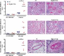
          It is controversial whether virus infections can contribute to the development of autoimmune diseases. Type I interferons (IFNs) are critical antiviral cytokines during virus infections and have also been implicated in the pathogenesis of systemic lupus erythematosus. Type I IFN is mainly produced by plasmacytoid dendritic cells (pDCs). The secretion of type I IFN of pDCs is modulated by Siglec-H, a DAP12-associated receptor on pDCs. In this study, we show that Siglec-H–deficient pDCs produce more of the type I IFN, IFN-α, in vitro and that Siglec-H knockout (KO) mice produce more IFN-α after murine cytomegalovirus (mCMV) infection in vivo. This did not impact control of viral replication. Remarkably, several weeks after a single mCMV infection, Siglec-H KO mice developed a severe form of systemic lupus–like autoimmune disease with strong kidney nephritis. In contrast, uninfected aging Siglec-H KO mice developed a mild form of systemic autoimmunity. The induction of systemic autoimmune disease after virus infection in Siglec-H KO mice was accompanied by a type I IFN signature and fully dependent on type I IFN signaling. These results show that Siglec-H normally serves as a modulator of type I IFN responses after infection with a persistent virus and thereby prevents induction of autoimmune disease.

          Related collections

          Most cited references53

          • Record: found
          • Abstract: found
          • Article: not found

          Interferon-inducible gene expression signature in peripheral blood cells of patients with severe lupus.

          Systemic lupus erythematosus (SLE) is a complex, inflammatory autoimmune disease that affects multiple organ systems. We used global gene expression profiling of peripheral blood mononuclear cells to identify distinct patterns of gene expression that distinguish most SLE patients from healthy controls. Strikingly, about half of the patients studied showed dysregulated expression of genes in the IFN pathway. Furthermore, this IFN gene expression "signature" served as a marker for more severe disease involving the kidneys, hematopoetic cells, and/or the central nervous system. These results provide insights into the genetic pathways underlying SLE, and identify a subgroup of patients who may benefit from therapies targeting the IFN pathway.
            Bookmark
            • Record: found
            • Abstract: found
            • Article: not found

            Plasmacytoid dendritic cells in immunity.

            Human and mouse plasmacytoid dendritic cells have been shown to correspond to a specialized cell population that produces large amounts of type I interferons in response to viruses, the so-called natural interferon-producing cells. As a result, intensive investigation is now focused on the potential functions of plasmacytoid dendritic cells in both innate and adaptive immunity. Here we review recent progress on the characterization of plasmacytoid dendritic cell origin, development, migration and function in immunity and tolerance, as well as their effect on human diseases.
              Bookmark
              • Record: found
              • Abstract: found
              • Article: not found

              Identification of cDC1- and cDC2-committed DC progenitors reveals early lineage priming at the common DC progenitor stage in the bone marrow.

              Mouse conventional dendritic cells (cDCs) can be classified into two functionally distinct lineages: the CD8α(+) (CD103(+)) cDC1 lineage, and the CD11b(+) cDC2 lineage. cDCs arise from a cascade of bone marrow (BM) DC-committed progenitor cells that include the common DC progenitors (CDPs) and pre-DCs, which exit the BM and seed peripheral tissues before differentiating locally into mature cDCs. Where and when commitment to the cDC1 or cDC2 lineage occurs remains poorly understood. Here we found that transcriptional signatures of the cDC1 and cDC2 lineages became evident at the single-cell level from the CDP stage. We also identified Siglec-H and Ly6C as lineage markers that distinguished pre-DC subpopulations committed to the cDC1 lineage (Siglec-H(-)Ly6C(-) pre-DCs) or cDC2 lineage (Siglec-H(-)Ly6C(+) pre-DCs). Our results indicate that commitment to the cDC1 or cDC2 lineage occurs in the BM and not in the periphery.
                Bookmark

                Author and article information

                Journal
                J Exp Med
                J. Exp. Med
                jem
                jem
                The Journal of Experimental Medicine
                The Rockefeller University Press
                0022-1007
                1540-9538
                25 July 2016
                : 213
                : 8
                : 1627-1644
                Affiliations
                [1 ]Division of Genetics, Department of Biology, University of Erlangen-Nürnberg, 91054 Erlangen, Germany
                [2 ]Nikolaus-Fiebiger-Zentrum, Division of Genetics, Department of Biology, University of Erlangen-Nürnberg, 91054 Erlangen, Germany
                [3 ]Division of Biochemistry, Department of Biology, University of Erlangen-Nürnberg, 91054 Erlangen, Germany
                [4 ]Department of Nephropathology, University of Erlangen-Nürnberg, 91054 Erlangen, Germany
                Author notes
                Correspondence to Lars Nitschke: lars.nitschke@ 123456fau.de ; or Thomas H. Winkler: thomas.winkler@ 123456fau.de
                [*]

                T.H. Winkler and L. Nitschke contributed equally to this paper.

                Author information
                http://orcid.org/0000-0002-4584-2733
                http://orcid.org/0000-0002-9870-3534
                http://orcid.org/0000-0002-9185-6255
                http://orcid.org/0000-0002-6803-4755
                Article
                20160189
                10.1084/jem.20160189
                4986536
                27377589
                dde16923-9301-4b4c-bb6a-8ca9435f04d6
                © 2016 Schmitt et al.

                This article is distributed under the terms of an Attribution–Noncommercial–Share Alike–No Mirror Sites license for the first six months after the publication date (see http://www.rupress.org/terms). After six months it is available under a Creative Commons License (Attribution–Noncommercial–Share Alike 3.0 Unported license, as described at http://creativecommons.org/licenses/by-nc-sa/3.0/).

                History
                : 08 February 2016
                : 13 May 2016
                Funding
                Funded by: Deutsche Forschungsgemeinschaft http://dx.doi.org/10.13039/501100001659
                Award ID: GRK1660
                Award ID: TRR130
                Award ID: CRC 1181
                Award ID: GRK1660
                Award ID: TRR130
                Categories
                Research Articles
                Article

                Medicine
                Medicine

                Comments

                Comment on this article