2
views
0
recommends
+1 Recommend
0 collections
    0
    shares
      • Record: found
      • Abstract: found
      • Article: found
      Is Open Access

      Correlation of radiological features of white epidermoid cysts with histopathological findings

      research-article

      Read this article at

      Bookmark
          There is no author summary for this article yet. Authors can add summaries to their articles on ScienceOpen to make them more accessible to a non-specialist audience.

          Abstract

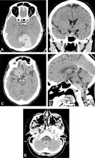
          Epidermoid cysts are benign congenital extra-axial lesions commonly found in the posterior fossa. These lesions have a characteristic imaging appearance on computed tomography (CT) scan and magnetic resonance imaging (MRI), but occasionally they may exhibit atypical radiological features, showing unusual hyperintensity on T1-weighted images (T1WI). Currently, such atypical appearance is referred to as white epidermoid. We present the imaging features of 5 cases of white epidermoid cyst and discuss the possible underlying etiology of this unusual radiological appearance. We retrospectively searched our electronic radiology database from January 2005 to December 2015 for all intracranial epidermoid cysts, which were confirmed either by typical MRI appearance or histopathological examination. All white epidermoid cases were evaluated with non-enhanced CT scan and multisequential MRI. Histopathological correlation was carried out in four white epidermoid cases. A total of 61 patients with epidermoid cyst were found, of those 5 (8%) were considered white epidermoids. These consisted of 3 females and 2 males, ranging in age between 31–63 years (average age was 51.8 years). Three patients had lesions located in the posterior fossa. The 2 other patients had lesions in the suprasellar region, with extension to the right middle cranial fossa in one. All 5 lesions were hyperdense on CT scan and hyperintense on T1WI. One patient demonstrated evidence of transformation of a classic epidermoid to a white epidermoid after partial resection. Histopathologically, cholesterol clefts were seen in 3 epidermoid cysts, each which also showed microcalcifications, proteinaceous material or melanin. Hemorrhage was demonstrated in one additional lesion. White epidermoid cyst is an unusual intracranial lesion that should be considered when encountered with an extra-axial T1 hyperintense lesion. The cause of this hyperintensity is not clearly understood, but the presence of cholesterol, microcalcifications, proteinaceous content and rarely hemorrhage or melanin may be contributing factors.

          Related collections

          Most cited references36

          • Record: found
          • Abstract: found
          • Article: not found

          Intracranial lesions with high signal intensity on T1-weighted MR images: differential diagnosis.

          Various substances, including methemoglobin, melanin, lipid, protein, calcium, iron, copper, and manganese, are responsible for the intrinsically high signal intensity observed in intracranial lesions at T1-weighted magnetic resonance (MR) imaging. Many of these substances have physical properties that lead to other specific imaging features as well. For example, lipid-containing lesions frequently produce chemical shift artifact, and some melanin-containing lesions exhibit a combination of high signal intensity on T1-weighted images and low signal intensity on T2-weighted images. The location and extent of a region of abnormal signal hyperintensity may be helpful for identifying rare diseases such as an ectopic posterior pituitary gland near the floor of the third ventricle, bilateral involvement of the dentate and lentiform nuclei in Cockayne syndrome, and involvement of the anterior temporal lobe and cerebellum in neurocutaneous melanosis. In cases in which diagnostically specific T1-weighted imaging features are lacking, findings obtained with other MR pulse sequences and other modalities can help narrow the differential diagnosis: An elevated glutamine or glutamate level at MR spectroscopy is suggestive of hepatic encephalopathy; a popcorn ball-like appearance at T2-weighted imaging, of cavernous malformations; and hyperattenuation at computed tomography, of mineral deposition disease. In many cases, a comparison of imaging features with clinical measures enables a specific diagnosis. © RSNA, 2012.
            Bookmark
            • Record: found
            • Abstract: found
            • Article: not found

            Intracranial epidermoid cyst with hemorrhage: MR imaging findings.

            We present a case of a 28-year-old woman with a cerebellopontine angle and prepontine cistern epidermoid cyst with unusual signal intensity. She presented with cranial nerve neuropathy and unsteady gait. MR imaging showed a tumor mass with central area of hemorrhage and a focal area of heterogeneous signal intensity with spotty enhancement, which correlated histologically to old blood in a cystic lumen and granulation of a cystic wall, with a large area of hemorrhage and mild vascularity.
              Bookmark
              • Record: found
              • Abstract: found
              • Article: not found

              Clinical, radiological, and pathological features of 24 atypical intracranial epidermoid cysts.

              Most intracranial epidermoid cysts typically present with long T1 and T2 signals on MR images. Other epidermoid cysts with atypical MR images are often misdiagnosed as other diseases. In this study the authors aimed to analyze the incidence and the clinical, radiological, and pathological features of atypical epidermoid cysts.
                Bookmark

                Author and article information

                Contributors
                djamjoom@ksu.edu.sa
                Journal
                Sci Rep
                Sci Rep
                Scientific Reports
                Nature Publishing Group UK (London )
                2045-2322
                10 February 2022
                10 February 2022
                2022
                : 12
                : 2314
                Affiliations
                [1 ]GRID grid.56302.32, ISNI 0000 0004 1773 5396, Department of Radiology and Medical Imaging, College of Medicine, , King Saud University, ; P.O. Box 7805, Riyadh, 11472 Saudi Arabia
                [2 ]GRID grid.412602.3, ISNI 0000 0000 9421 8094, Department of Radiology, College of Medicine, , Qassim University, ; Buraidah, Saudi Arabia
                [3 ]GRID grid.410356.5, ISNI 0000 0004 1936 8331, Kingston Health Sciences Centre, , Queen’s University, ; Kingston, Canada
                Article
                6167
                10.1038/s41598-022-06167-x
                8831518
                35145173
                def675c8-cc78-4a6d-9b93-46306789544b
                © The Author(s) 2022

                Open Access This article is licensed under a Creative Commons Attribution 4.0 International License, which permits use, sharing, adaptation, distribution and reproduction in any medium or format, as long as you give appropriate credit to the original author(s) and the source, provide a link to the Creative Commons licence, and indicate if changes were made. The images or other third party material in this article are included in the article's Creative Commons licence, unless indicated otherwise in a credit line to the material. If material is not included in the article's Creative Commons licence and your intended use is not permitted by statutory regulation or exceeds the permitted use, you will need to obtain permission directly from the copyright holder. To view a copy of this licence, visit http://creativecommons.org/licenses/by/4.0/.

                History
                : 30 August 2021
                : 25 January 2022
                Categories
                Article
                Custom metadata
                © The Author(s) 2022

                Uncategorized
                neurological disorders,diseases of the nervous system
                Uncategorized
                neurological disorders, diseases of the nervous system

                Comments

                Comment on this article