3
views
0
recommends
+1 Recommend
1 collections
    0
    shares
      • Record: found
      • Abstract: found
      • Article: not found

      Incidental CT findings in the lungs in COVID-19 patients presenting with abdominal pain

      case-report

      Read this article at

      ScienceOpenPublisherPMC
      Bookmark
          There is no author summary for this article yet. Authors can add summaries to their articles on ScienceOpen to make them more accessible to a non-specialist audience.

          Abstract

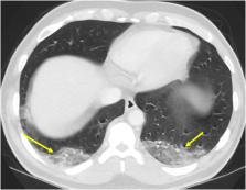
          As the 2019 novel coronavirus disease (COVID-19) continues to spread, some patients are presenting with abdominal symptoms without respiratory complaints. Our case series documents four patients who presented with abdominal symptoms whose abdominopelvic CT revealed incidental pulmonary parenchymal findings in the imaged lung bases and were subsequently confirmed positive for COVID-19 via laboratory testing. It remains to be seen whether these patients will eventually develop respiratory symptoms. While it is possible that the patients' abdominal complaints are coincidental with CT findings, it is interesting that patients can have such extensive incidental disease in the lungs on CT without respiratory symptoms.

          Highlights

          • Abdominal pain is being more recognized as a potential presenting for COVID-19.

          • Lung findings in COVID-19 patients may not correlate to respiratory symptoms.

          • Noting atypical presentation of COVID-19 allows for earlier quarantine and testing.

          • Early identification can further limit spread of COVID-19.

          Related collections

          Most cited references11

          • Record: found
          • Abstract: found
          • Article: not found

          CT Imaging Features of 2019 Novel Coronavirus (2019-nCoV)

          In this retrospective case series, chest CT scans of 21 symptomatic patients from China infected with the 2019 novel coronavirus (2019-nCoV) were reviewed, with emphasis on identifying and characterizing the most common findings. Typical CT findings included bilateral pulmonary parenchymal ground-glass and consolidative pulmonary opacities, sometimes with a rounded morphology and a peripheral lung distribution. Notably, lung cavitation, discrete pulmonary nodules, pleural effusions, and lymphadenopathy were absent. Follow-up imaging in a subset of patients during the study time window often demonstrated mild or moderate progression of disease, as manifested by increasing extent and density of lung opacities. © RSNA, 2020
            Bookmark
            • Record: found
            • Abstract: found
            • Article: not found

            Chest CT Findings in Coronavirus Disease-19 (COVID-19): Relationship to Duration of Infection

            Abstract In this retrospective study, chest CTs of 121 symptomatic patients infected with coronavirus disease-19 (COVID-19) from four centers in China from January 18, 2020 to February 2, 2020 were reviewed for common CT findings in relationship to the time between symptom onset and the initial CT scan (i.e. early, 0-2 days (36 patients), intermediate 3-5 days (33 patients), late 6-12 days (25 patients)). The hallmarks of COVID-19 infection on imaging were bilateral and peripheral ground-glass and consolidative pulmonary opacities. Notably, 20/36 (56%) of early patients had a normal CT. With a longer time after the onset of symptoms, CT findings were more frequent, including consolidation, bilateral and peripheral disease, greater total lung involvement, linear opacities, “crazy-paving” pattern and the “reverse halo” sign. Bilateral lung involvement was observed in 10/36 early patients (28%), 25/33 intermediate patients (76%), and 22/25 late patients (88%).
              Bookmark
              • Record: found
              • Abstract: found
              • Article: not found

              Clinical characteristics and imaging manifestations of the 2019 novel coronavirus disease (COVID-19):A multi-center study in Wenzhou city, Zhejiang, China

              Background Little is known about COVID-19 outside Hubei. The aim of this paper was to describe the clinical characteristics and imaging manifestations of hospitalized patients with confirmed COVID-19 infection in Wenzhou, Zhejiang, China. Methods In this retrospective cohort study, 149 RT-PCR confirmed positive patients were consecutively enrolled from January 17th to February 10th, 2020 in three tertiary hospitals of Wenzhou. Outcomes were followed up until Feb 15th, 2020. Findings A total of 85 patients had Hubei travel/residence history, while another 49 had contact with people from Hubei and 15 had no traceable exposure history to Hubei. Fever, cough and expectoration were the most common symptoms, 14 patients had decreased oxygen saturation, 33 had leukopenia, 53 had lymphopenia, and 82 had elevated C-reactive protein. On chest computed tomography (CT), lung segments 6 and 10 were mostly involved. A total of 287 segments presented ground glass opacity, 637 presented mixed opacity and 170 presented consolidation. Lesions were more localized in the peripheral lung with a patchy form. No significant difference was found between patients with or without Hubei exposure history. Seventeen patients had normal CT on admission of these, 12 had negative findings even10 days later. Interpretation Most patients presented with a mild infection in our study. The imaging pattern of multifocal peripheral ground glass or mixed opacity with predominance in the lower lung is highly suspicious of COVID-19 in the first week of disease onset. Nevetheless, some patients can present with a normal chest finding despite testing positive for COVID-19. Funding: We did not receive any fundings.
                Bookmark

                Author and article information

                Contributors
                Journal
                Clin Imaging
                Clin Imaging
                Clinical Imaging
                Published by Elsevier Inc.
                0899-7071
                1873-4499
                28 May 2020
                28 May 2020
                Affiliations
                Icahn School of Medicine, Department of Diagnostic, Molecular, and Interventional Radiology, 1 Gustave Levy Place, Box 1234, NY, New York 10029, United States of America
                Author notes
                [* ]Corresponding author at: 1 Gustave Levy Place, Box 1234, NY, New York 10029, United States of America. nicholas.voutsinas@ 123456mountsinai.org
                Article
                S0899-7071(20)30190-X
                10.1016/j.clinimag.2020.05.021
                7255745
                df0be34a-2f24-4d2c-859c-c91f0359a05a
                © 2020 Published by Elsevier Inc.

                Since January 2020 Elsevier has created a COVID-19 resource centre with free information in English and Mandarin on the novel coronavirus COVID-19. The COVID-19 resource centre is hosted on Elsevier Connect, the company's public news and information website. Elsevier hereby grants permission to make all its COVID-19-related research that is available on the COVID-19 resource centre - including this research content - immediately available in PubMed Central and other publicly funded repositories, such as the WHO COVID database with rights for unrestricted research re-use and analyses in any form or by any means with acknowledgement of the original source. These permissions are granted for free by Elsevier for as long as the COVID-19 resource centre remains active.

                History
                : 10 April 2020
                : 9 May 2020
                : 27 May 2020
                Categories
                Article

                covid-19,abdominal pain,viral pneumonia,ct abdomen/pelvis,lung bases

                Comments

                Comment on this article