2
views
0
recommends
+1 Recommend
0 collections
    0
    shares
      • Record: found
      • Abstract: found
      • Article: found
      Is Open Access

      Incidental Finding of Secondary Tumoral Calcinosis Following Cardiothoracic Surgery: The Role of Multimodality Imaging Including Spectral Detector Computed Tomography

      case-report

      Read this article at

      Bookmark
          There is no author summary for this article yet. Authors can add summaries to their articles on ScienceOpen to make them more accessible to a non-specialist audience.

          Abstract

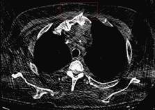
          Tumoral calcinosis is a rare syndrome that affects mostly soft tissues. It is characterized by calcium salt deposition in the periarticular soft tissue surrounding bony structures forming slow-growing, seldom asymptomatic masses. This case report describes a 41-year-old male with end-stage renal disease on home hemodialysis, who presented with an unusual rapidly progressive mass overlying the manubrium and suprasternal notch, following recent cardiothoracic surgery, which was initially felt to be a hematoma. The case highlights the role of spectral detector computed tomography (SDCT) in reaching the correct diagnosis of tumoral calcinosis as well as demonstrating additional changes of ectopic parathyroid hyperplasia in the anterior mediastinum.

          Related collections

          Most cited references14

          • Record: found
          • Abstract: found
          • Article: not found

          Review of tumoral calcinosis: A rare clinico-pathological entity.

          Tumoral calcinosis (TC) has long been a controversial clinico-pathological entity. Its pathogenesis and genetic background have been gradually unravelled since its first description in 1943. According to the presence or absence of an underlying calcifying disease process, TC has been divided into primary and secondary varieties. Two subtypes of the primary variety exist; a hyper-phosphatemic type with familial basis represented by mutations in GalNAc transferase 3 gene (GALNT3), KLOTHO or Fibroblast growth factor 23 (FGF23) genes, and a normo-phosphatemic type with growing evidence of underlying familial base represented by mutation in SAMD9 gene. The secondary variety is mainly associated with chronic renal failure and the resulting secondary or tertiary hyperparathyroidism. Diagnosis of TC relies on typical radiographic features (on plain radiographs and computed tomography) and the biochemical profile. Magnetic resonance imaging can be done in difficult cases, and scintigraphy reflects the disease activity. Treatment is mainly surgical for the primary variety; however, a stage-oriented conservative approach using phosphate binders, phosphate restricted diets and acetazolamide should be considered before the surgical approach is pursued due to the high rate of recurrences and complications after surgical intervention. Medical treatment is the mainstay for treatment of the secondary variety, with failure warranting subtotal or total parathyroidectomy. Surgical intervention in these patients should be kept as a last resort.
            Bookmark
            • Record: found
            • Abstract: found
            • Article: not found

            Tumoral calcinosis: pearls, polemics, and alternative possibilities.

            Massive periarticular calcinosis of the soft tissues is a unique but not rare radiographic finding. On the contrary, tumoral calcinosis is a rare familial disease. Unfortunately, the term tumoral calcinosis has been liberally and imprecisely used to describe any massive collection of periarticular calcification, although this term actually refers to a hereditary condition associated with massive periarticular calcification. The inconsistent use of this term has created confusion throughout the literature. More important, if the radiologist is unfamiliar with tumoral calcinosis or disease processes that mimic this condition, then diagnosis could be impeded, treatment could be delayed, and undue alarm could be raised, possibly leading to unwarranted surgical procedures. The soft-tissue lesions of tumoral calcinosis are typically lobulated, well-demarcated calcifications that are most often distributed along the extensor surfaces of large joints. There are many conditions with similar appearances, including the calcinosis of chronic renal failure, calcinosis universalis, calcinosis circumscripta, calcific tendonitis, synovial osteochondromatosis, synovial sarcoma, osteosarcoma, myositis ossificans, tophaceous gout, and calcific myonecrosis. The radiologist plays a critical role in avoiding unnecessary invasive procedures and in guiding the selection of appropriate tests that can result in a conclusive diagnosis of tumoral calcinosis. Copyright RSNA, 2006.
              Bookmark
              • Record: found
              • Abstract: not found
              • Article: not found

              TUMORAL CALCINOSIS

                Bookmark

                Author and article information

                Journal
                Cureus
                Cureus
                2168-8184
                Cureus
                Cureus (Palo Alto (CA) )
                2168-8184
                16 July 2022
                July 2022
                : 14
                : 7
                : e26929
                Affiliations
                [1 ] Medicine, "Carol Davila" University of Medicine and Pharmacy, Bucharest, ROU
                [2 ] Hematology, Center of Hematology and Bone Marrow Transplantation, Fundeni Clinical Institute, Bucharest, ROU
                [3 ] Cardiovascular Medicine, Baylor College of Medicine, Houston, USA
                [4 ] Cardiovascular Medicine, Georgetown University School of Medicine, MedStar Washington Hospital Center, Washington, USA
                [5 ] Radiology, University Hospitals Cleveland Medical Center, Cleveland, USA
                [6 ] Diagnostic Radiology, Case Western Reserve University School of Medicine, Cleveland, USA
                Author notes
                Mihnea-Alexandru Găman mihneagaman@ 123456yahoo.com
                Article
                10.7759/cureus.26929
                9379215
                e0dc11cd-3289-443a-b3bf-4e27321d6e1e
                Copyright © 2022, Găman et al.

                This is an open access article distributed under the terms of the Creative Commons Attribution License, which permits unrestricted use, distribution, and reproduction in any medium, provided the original author and source are credited.

                History
                : 16 July 2022
                Categories
                Endocrinology/Diabetes/Metabolism
                Internal Medicine
                Nephrology

                spectral detector computed tomography,end-stage renal disease,renal failure,hyperparathyroidism,tumoral calcinosis

                Comments

                Comment on this article