6
views
0
recommends
+1 Recommend
0 collections
    0
    shares
      • Record: found
      • Abstract: found
      • Article: found
      Is Open Access

      [ 18F]fallypride-PET/CT Analysis of the Dopamine D 2/D 3 Receptor in the Hemiparkinsonian Rat Brain Following Intrastriatal Botulinum Neurotoxin A Injection

      research-article

      Read this article at

      Bookmark
          There is no author summary for this article yet. Authors can add summaries to their articles on ScienceOpen to make them more accessible to a non-specialist audience.

          Abstract

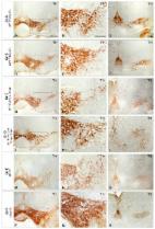
          Intrastriatal injection of botulinum neurotoxin A (BoNT-A) results in improved motor behavior of hemiparkinsonian (hemi-PD) rats, an animal model for Parkinson’s disease. The caudate–putamen (CPu), as the main input nucleus of the basal ganglia loop, is fundamentally involved in motor function and directly interacts with the dopaminergic system. To determine receptor-mediated explanations for the BoNT-A effect, we analyzed the dopamine D 2/D 3 receptor (D 2/D 3R) in the CPu of 6-hydroxydopamine (6-OHDA)-induced hemi-PD rats by [ 18F]fallypride-PET/CT scans one, three, and six months post-BoNT-A or -sham-BoNT-A injection. Male Wistar rats were assigned to three different groups: controls, sham-injected hemi-PD rats, and BoNT-A-injected hemi-PD rats. Disease-specific motor impairment was verified by apomorphine and amphetamine rotation testing. Animal-specific magnetic resonance imaging was performed for co-registration and anatomical reference. PET quantification was achieved using PMOD software with the simplified reference tissue model 2. Hemi-PD rats exhibited a constant increase of 23% in D 2/D 3R availability in the CPu, which was almost normalized by intrastriatal application of BoNT-A. Importantly, the BoNT-A effect on striatal D 2/D 3R significantly correlated with behavioral results in the apomorphine rotation test. Our results suggest a therapeutic effect of BoNT-A on the impaired motor behavior of hemi-PD rats by reducing interhemispheric changes of striatal D 2/D 3R.

          Related collections

          Most cited references73

          • Record: found
          • Abstract: not found
          • Article: not found

          Quantitative recording of rotational behavior in rats after 6-hydroxy-dopamine lesions of the nigrostriatal dopamine system.

            Bookmark
            • Record: found
            • Abstract: found
            • Article: found
            Is Open Access

            Parkinson’s Disease: From Pathogenesis to Pharmacogenomics

            Parkinson’s disease (PD) is the second most important age-related neurodegenerative disorder in developed societies, after Alzheimer’s disease, with a prevalence ranging from 41 per 100,000 in the fourth decade of life to over 1900 per 100,000 in people over 80 years of age. As a movement disorder, the PD phenotype is characterized by rigidity, resting tremor, and bradykinesia. Parkinson’s disease -related neurodegeneration is likely to occur several decades before the onset of the motor symptoms. Potential risk factors include environmental toxins, drugs, pesticides, brain microtrauma, focal cerebrovascular damage, and genomic defects. Parkinson’s disease neuropathology is characterized by a selective loss of dopaminergic neurons in the substantia nigra pars compacta, with widespread involvement of other central nervous system (CNS) structures and peripheral tissues. Pathogenic mechanisms associated with genomic, epigenetic and environmental factors lead to conformational changes and deposits of key proteins due to abnormalities in the ubiquitin–proteasome system together with dysregulation of mitochondrial function and oxidative stress. Conventional pharmacological treatments for PD are dopamine precursors (levodopa, l-DOPA, l-3,4 dihidroxifenilalanina), and other symptomatic treatments including dopamine agonists (amantadine, apomorphine, bromocriptine, cabergoline, lisuride, pergolide, pramipexole, ropinirole, rotigotine), monoamine oxidase (MAO) inhibitors (selegiline, rasagiline), and catechol-O-methyltransferase (COMT) inhibitors (entacapone, tolcapone). The chronic administration of antiparkinsonian drugs currently induces the “wearing-off phenomenon”, with additional psychomotor and autonomic complications. In order to minimize these clinical complications, novel compounds have been developed. Novel drugs and bioproducts for the treatment of PD should address dopaminergic neuroprotection to reduce premature neurodegeneration in addition to enhancing dopaminergic neurotransmission. Since biochemical changes and therapeutic outcomes are highly dependent upon the genomic profiles of PD patients, personalized treatments should rely on pharmacogenetic procedures to optimize therapeutics.
              Bookmark
              • Record: found
              • Abstract: found
              • Article: not found

              Neurotoxin-based models of Parkinson's disease.

              Animal experimentation in the Parkinson's disease (PD) field is a classic example of how the use of animal models to study diseases can have a significant impact on human health. Among the different neurotoxin-based animal models of PD that are presently available, the 6-hydroxydopamine (6-OHDA) and the 1-methyl-4-phenyl-1,2,3,6-tetrahydropyridine (MPTP) models have been established and validated as useful models for the development of therapeutic strategies aimed to treat motor symptoms and to study alterations of the basal ganglia that occur in this disease. The 6-OHDA rat model and the MPTP primate model have contributed enormously to translate animal experimentation into clinical practice, including pharmacological treatments and deep brain stimulation of the subthalamic nucleus. These models, along with the MPTP mouse model, are helping to elucidate the pathogenic mechanism of neurodegeneration in PD. The roles of oxidative stress, apoptosis, mitochondrial dysfunction, inflammation, and impairment of the protein degradation pathways have also come under careful consideration thanks to these models. The more recently developed paraquat and rotenone rodent models are also contributing to our understanding of neuronal cell death. However, none of the neuroprotective strategies that have worked in the pre-clinical stage have thus far been successfully translated to a clinical setting to treat PD patients. At the same time, the lack of any effective neuroprotective strategy for PD is preventing the validation of any one particular model as a screening tool for such neuroprotective strategies. Therefore, it seems that we are trapped in a vicious circle that casts doubt on the suitability of the neurotoxin-based models for this purpose. Here, we discuss how epidemiological data may help to validate a specific model with data linking a lower risk of developing PD with nutritional/consumption habits or with a specific chronic drug therapy. Copyright © 2012 IBRO. Published by Elsevier Ltd. All rights reserved.
                Bookmark

                Author and article information

                Journal
                Molecules
                Molecules
                molecules
                Molecules : A Journal of Synthetic Chemistry and Natural Product Chemistry
                MDPI
                1420-3049
                06 March 2018
                March 2018
                : 23
                : 3
                : 587
                Affiliations
                [1 ]Institute of Anatomy, Rostock University Medical Center, Gertrudenstrasse 9, 18057 Rostock, Germany; alexander.hawlitschka@ 123456med.uni-rostock.de (A.H.); andreas.wree@ 123456med.uni-rostock.de (A.W.)
                [2 ]Department of Nuclear Medicine, Rostock University Medical Centre, Gertrudenplatz 1, 18057 Rostock, Germany; jens.kurth@ 123456med.uni-rostock.de (J.K.); alexander.hohn@ 123456med.uni-rostock.de (A.H.); bernd.krause@ 123456med.uni-rostock.de (B.J.K.)
                [3 ]Core Facility Multimodal Small Animal Imaging, Rostock University Medical Center, Schillingallee 69a, 18057 Rostock, Germany; Jan.Stenzel@ 123456med.uni-rostock.de (J.S.); tobias.lindner@ 123456med.uni-rostock.de (T.L.); stefan.polei@ 123456gmx.de (S.P.)
                Author notes
                [* ]Correspondence: teresa.mann@ 123456med.uni-rostock.de ; Tel.: +49-381-494-8433
                Author information
                https://orcid.org/0000-0001-6864-4513
                https://orcid.org/0000-0001-7826-3132
                Article
                molecules-23-00587
                10.3390/molecules23030587
                6017015
                29509680
                e266996c-f1fe-4182-b9d7-3967dbaa56cd
                © 2018 by the authors.

                Licensee MDPI, Basel, Switzerland. This article is an open access article distributed under the terms and conditions of the Creative Commons Attribution (CC BY) license ( http://creativecommons.org/licenses/by/4.0/).

                History
                : 30 January 2018
                : 04 March 2018
                Categories
                Article

                d2/d3 receptors,hemiparkinsonian rat model,botulinum neurotoxin a,basal ganglia,striatum,parkinson’s disease,small animal imaging,pet/ct,[18f]fallypride,mri

                Comments

                Comment on this article