22
views
0
recommends
+1 Recommend
0 collections
    0
    shares
      • Record: found
      • Abstract: found
      • Article: found
      Is Open Access

      Targeting protein tyrosine phosphatase σ after myocardial infarction restores cardiac sympathetic innervation and prevents arrhythmias

      research-article

      Read this article at

      Bookmark
          There is no author summary for this article yet. Authors can add summaries to their articles on ScienceOpen to make them more accessible to a non-specialist audience.

          Abstract

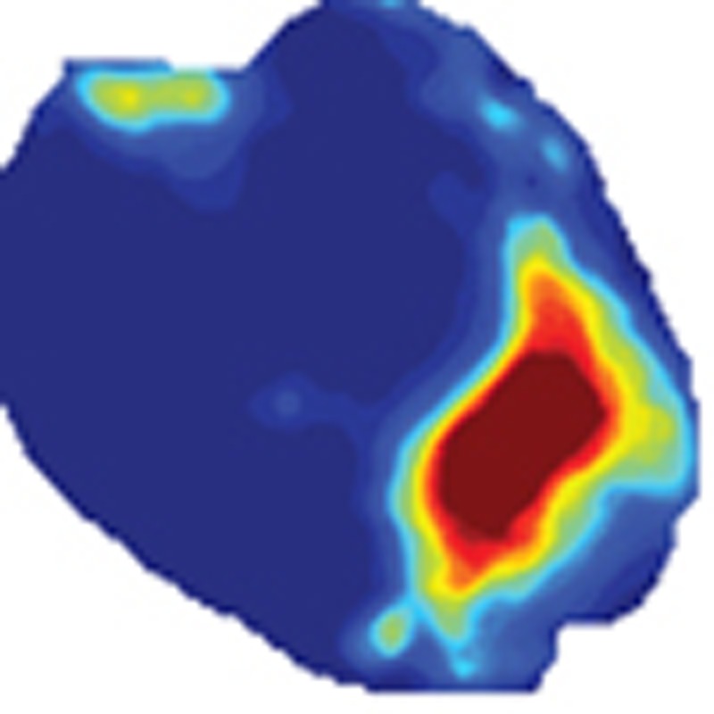
          Millions of people suffer a myocardial infarction (MI) every year, and those who survive have increased risk of arrhythmias and sudden cardiac death. Recent clinical studies have identified sympathetic denervation as a predictor of increased arrhythmia susceptibility. Chondroitin sulfate proteoglycans present in the cardiac scar after MI prevent sympathetic reinnervation by binding the neuronal protein tyrosine phosphatase receptor σ (PTPσ). Here we show that the absence of PTPσ, or pharmacologic modulation of PTPσ by the novel intracellular sigma peptide (ISP) beginning 3 days after injury, restores sympathetic innervation to the scar and markedly reduces arrhythmia susceptibility. Using optical mapping we observe increased dispersion of action potential duration, supersensitivity to β-adrenergic receptor stimulation and Ca 2+ mishandling following MI. Sympathetic reinnervation prevents these changes and renders hearts remarkably resistant to induced arrhythmias.

          Abstract

          Chondroitin sulfate proteoglycans (CSP) in the myocardial scar inhibit the tissue’s reinnervation, rendering it prone to arrhythmia. Here the authors show that blocking the activity of the CSP receptor, protein tyrosine phosphatase receptor σ, promotes scar reinnervation and prevents arrhythmia in mice.

          Related collections

          Most cited references62

          • Record: found
          • Abstract: found
          • Article: not found

          Prophylactic implantation of a defibrillator in patients with myocardial infarction and reduced ejection fraction.

          Patients with reduced left ventricular function after myocardial infarction are at risk for life-threatening ventricular arrhythmias. This randomized trial was designed to evaluate the effect of an implantable defibrillator on survival in such patients. Over the course of four years, we enrolled 1232 patients with a prior myocardial infarction and a left ventricular ejection fraction of 0.30 or less. Patients were randomly assigned in a 3:2 ratio to receive an implantable defibrillator (742 patients) or conventional medical therapy (490 patients). Invasive electrophysiological testing for risk stratification was not required. Death from any cause was the end point. The clinical characteristics at base line and the prevalence of medication use at the time of the last follow-up visit were similar in the two treatment groups. During an average follow-up of 20 months, the mortality rates were 19.8 percent in the conventional-therapy group and 14.2 percent in the defibrillator group. The hazard ratio for the risk of death from any cause in the defibrillator group as compared with the conventional-therapy group was 0.69 (95 percent confidence interval, 0.51 to 0.93; P=0.016). The effect of defibrillator therapy on survival was similar in subgroup analyses stratified according to age, sex, ejection fraction, New York Heart Association class, and the QRS interval. In patients with a prior myocardial infarction and advanced left ventricular dysfunction, prophylactic implantation of a defibrillator improves survival and should be considered as a recommended therapy.
            Bookmark
            • Record: found
            • Abstract: found
            • Article: not found

            Diabetic Hyperglycemia activates CaMKII and Arrhythmias by O linked Glycosylation

            Summary Ca2+-Calmodulin dependent protein kinase II (CaMKII) is a regulatory node in heart and brain, and its chronic activation can be pathological. CaMKII activation seen in heart failure can directly induce pathological changes in ion channels, Ca2+ handling and gene transcription. 1 Here we discover a novel mechanism linking CaMKII and hyperglycemic signaling in diabetes mellitus, which is a key risk factor for heart 2 and neurodegenerative diseases. 3,4 Acute hyperglycemia causes covalent modification of CaMKII by O-linked N-acetylglucosamine (O-GlcNAc). O-GlcNAc modification of CaMKII at Ser-279 activates CaMKII autonomously, creating molecular memory even after [Ca2+] declines. O-GlcNAc modified CaMKII is increased in heart and brain from diabetic humans and rats. In cardiomyocytes, increased [glucose] significantly enhances CaMKII-dependent activation of spontaneous sarcoplasmic reticulum (SR) Ca2+ release events that can contribute to cardiac mechanical dysfunction and arrhythmias. 1 These effects were prevented by pharmacological inhibition of O-GlcNAc signaling or genetic ablation of CaMKIIδ. In intact perfused hearts, arrhythmias were enhanced by increased [glucose] via O-GlcNAc-and CaMKII-dependent pathways. In diabetic animals, acute blockade of O-GlcNAc inhibited arrhythmogenesis. Thus, O-GlcNAc modification of CaMKII is a novel signaling event in pathways that may contribute critically to cardiac and neuronal pathophysiology in diabetes and other diseases.
              Bookmark
              • Record: found
              • Abstract: found
              • Article: not found

              Sudden death in patients with myocardial infarction and left ventricular dysfunction, heart failure, or both.

              The risk of sudden death from cardiac causes is increased among survivors of acute myocardial infarction with reduced left ventricular systolic function. We assessed the risk and time course of sudden death in high-risk patients after myocardial infarction. We studied 14,609 patients with left ventricular dysfunction, heart failure, or both after myocardial infarction to assess the incidence and timing of sudden unexpected death or cardiac arrest with resuscitation in relation to the left ventricular ejection fraction. Of 14,609 patients, 1067 (7 percent) had an event a median of 180 days after myocardial infarction: 903 died suddenly, and 164 were resuscitated after cardiac arrest. The risk was highest in the first 30 days after myocardial infarction--1.4 percent per month (95 percent confidence interval, 1.2 to 1.6 percent)--and decreased to 0.14 percent per month (95 percent confidence interval, 0.11 to 0.18 percent) after 2 years. Patients with a left ventricular ejection fraction of 30 percent or less were at highest risk in this early period (rate, 2.3 percent per month; 95 percent confidence interval, 1.8 to 2.8 percent). Nineteen percent of all sudden deaths or episodes of cardiac arrest with resuscitation occurred within the first 30 days after myocardial infarction, and 83 percent of all patients who died suddenly did so in the first 30 days after hospital discharge. Each decrease of 5 percentage points in the left ventricular ejection fraction was associated with a 21 percent adjusted increase in the risk of sudden death or cardiac arrest with resuscitation in the first 30 days. The risk of sudden death is highest in the first 30 days after myocardial infarction among patients with left ventricular dysfunction, heart failure, or both. Thus, earlier implementation of strategies for preventing sudden death may be warranted in selected patients. Copyright 2005 Massachusetts Medical Society.
                Bookmark

                Author and article information

                Journal
                Nat Commun
                Nat Commun
                Nature Communications
                Nature Pub. Group
                2041-1723
                02 February 2015
                : 6
                : 6235
                Affiliations
                [1 ]Department of Physiology and Pharmacology, Neuroscience Graduate Program, Oregon Health and Science University , Portland, Oregon 97239, USA
                [2 ]Department of Neurology, Oregon Health and Science University , Portland, Oregon 97239, USA
                [3 ]Department of Pharmacology, University of California , Davis, California 95616, USA
                [4 ]Department of Neurosciences, Case Western Reserve University , Cleveland, Ohio 44106, USA
                Author notes
                Article
                ncomms7235
                10.1038/ncomms7235
                4315356
                25639594
                e576711e-56d7-4dfe-a146-dc32d15f4f24
                Copyright © 2015, Nature Publishing Group, a division of Macmillan Publishers Limited. All Rights Reserved.

                This work is licensed under a Creative Commons Attribution 4.0 International License. The images or other third party material in this article are included in the article’s Creative Commons license, unless indicated otherwise in the credit line; if the material is not included under the Creative Commons license, users will need to obtain permission from the license holder to reproduce the material. To view a copy of this license, visit http://creativecommons.org/licenses/by/4.0/

                History
                : 06 November 2014
                : 07 January 2015
                Categories
                Article

                Uncategorized
                Uncategorized

                Comments

                Comment on this article