35
views
0
recommends
+1 Recommend
0 collections
    0
    shares
      • Record: found
      • Abstract: found
      • Article: not found

      Development and application of a Japanese model of the WHO fracture risk assessment tool (FRAX™)

      research-article

      Read this article at

      ScienceOpenPublisherPMC
      Bookmark
          There is no author summary for this article yet. Authors can add summaries to their articles on ScienceOpen to make them more accessible to a non-specialist audience.

          Abstract

          Summary

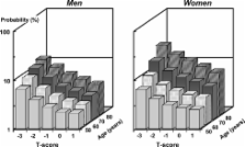
          The present study estimated the 10-year probability using the Japanese version of WHO fracture risk assessment tool (FRAX™) in order to determine fracture probabilities that correspond to intervention thresholds currently used in Japan and to resolve some issues for its use in Japan.

          Introduction

          The objective of the present study was to evaluate a Japanese version of the WHO fracture risk assessment (FRAX™) tool to compute 10-year probabilities of osteoporotic fracture in Japanese men and women. Since lumbar spine bone mineral density (BMD) is used preferentially as a site for assessment, and densitometers use Japanese reference data, a second aim was to investigate the suitability and impact of this practice in Japan.

          Methods

          Fracture probabilities were computed from published data on the fracture and death hazards in Japan. Probabilities took account of age, sex, the presence of clinical risk factors and femoral neck BMD. Fracture probabilities were determined that were equivalent to intervention thresholds currently used in Japan. The difference between T-scores derived from international reference data and that using Japanese-specific normal ranges was estimated from published sources. The gradient of risk of BMD for fracture in Japan was compared to that for BMD at the lumbar spine in the Hiroshima cohort.

          Results

          The 10-year probabilities of a major osteoporosis-related fracture that corresponded to current intervention thresholds ranged from approximately 5% at the age of 50 years to more than 20% at the age of 80 years. The use of femoral neck BMD predicts fracture as well as or better than BMD tests at the lumbar spine. There were small differences in T-scores between those used for the model and those derived from a Japanese reference population.

          Conclusions

          The FRAX™ tool has been used to determine possible thresholds for therapeutic intervention, based on equivalence of risk with current guidelines. The approach will need to be supported by appropriate health economic analyses. Femoral neck BMD is suitable for the prediction of fracture risk among Japanese. However, when applying the FRAX™ model to Japan, T-scores and Z-scores should be converted to those derived from the international reference.

          Related collections

          Most cited references16

          • Record: found
          • Abstract: found
          • Article: not found

          Predictive value of BMD for hip and other fractures.

          The relationship between BMD and fracture risk was estimated in a meta-analysis of data from 12 cohort studies of approximately 39,000 men and women. Low hip BMD was an important predictor of fracture risk. The prediction of hip fracture with hip BMD also depended on age and z score. The aim of this study was to quantify the relationship between BMD and fracture risk and examine the effect of age, sex, time since measurement, and initial BMD value. We studied 9891 men and 29,082 women from 12 cohorts comprising EVOS/EPOS, EPIDOS, OFELY, CaMos, Rochester, Sheffield, Rotterdam, Kuopio, DOES, Hiroshima, and 2 cohorts from Gothenburg. Cohorts were followed for up to 16.3 years and a total of 168,366 person-years. The effect of BMD on fracture risk was examined using a Poisson model in each cohort and each sex separately. Results of the different studies were then merged using weighted coefficients. BMD measurement at the femoral neck with DXA was a strong predictor of hip fractures both in men and women with a similar predictive ability. At the age of 65 years, risk ratio increased by 2.94 (95% CI = 2.02-4.27) in men and by 2.88 (95% CI = 2.31-3.59) in women for each SD decrease in BMD. However, the effect was dependent on age, with a significantly higher gradient of risk at age 50 years than at age 80 years. Although the gradient of hip fracture risk decreased with age, the absolute risk still rose markedly with age. For any fracture and for any osteoporotic fracture, the gradient of risk was lower than for hip fractures. At the age of 65 years, the risk of osteoporotic fractures increased in men by 1.41 per SD decrease in BMD (95% CI = 1.33-1.51) and in women by 1.38 per SD (95% CI = 1.28-1.48). In contrast with hip fracture risk, the gradient of risk increased with age. For the prediction of any osteoporotic fracture (and any fracture), there was a higher gradient of risk the lower the BMD. At a z score of -4 SD, the risk gradient was 2.10 per SD (95% CI = 1.63-2.71) and at a z score of -1 SD, the risk was 1.73 per SD (95% CI = 1.59-1.89) in men and women combined. A similar but less pronounced and nonsignificant effect was observed for hip fractures. Data for ultrasound and peripheral measurements were available from three cohorts. The predictive ability of these devices was somewhat less than that of DXA measurements at the femoral neck by age, sex, and BMD value. We conclude that BMD is a risk factor for fracture of substantial importance and is similar in both sexes. Its validation on an international basis permits its use in case finding strategies. Its use should, however, take account of the variations in predictive value with age and BMD.
            Bookmark
            • Record: found
            • Abstract: found
            • Article: not found

            A meta-analysis of previous fracture and subsequent fracture risk.

            Previous fracture is a well-documented risk factor for future fracture. The aim of this study was to quantify this risk on an international basis and to explore the relationship of this risk with age, sex, and bone mineral density (BMD). We studied 15259 men and 44902 women from 11 cohorts comprising EVOS/EPOS, OFELY, CaMos, Rochester, Sheffield, Rotterdam, Kuopio, DOES, Hiroshima, and two cohorts from Gothenburg. Cohorts were followed for a total of 250000 person-years. The effect of a prior history of fracture on the risk of any fracture, any osteoporotic fracture, and hip fracture alone was examined using a Poisson model for each sex from each cohort. Covariates examined were age, sex, and BMD. The results of the different studies were merged by using the weighted beta-coefficients. A previous fracture history was associated with a significantly increased risk of any fracture compared with individuals without a prior fracture (RR = 1.86; 95% CI = 1.75-1.98). The risk ratio was similar for the outcome of osteoporotic fracture or for hip fracture. There was no significant difference in risk ratio between men and women. Risk ratio (RR) was marginally downward adjusted when account was taken of BMD. Low BMD explained a minority of the risk for any fracture (8%) and for hip fracture (22%). The risk ratio was stable with age except in the case of hip fracture outcome where the risk ratio decreased significantly with age. We conclude that previous history of fracture confers an increased risk of fracture of substantial importance beyond that explained by measurement of BMD. Its validation on an international basis permits the use of this risk factor in case finding strategies.
              Bookmark
              • Record: found
              • Abstract: found
              • Article: not found

              Updated data on proximal femur bone mineral levels of US adults.

              This paper describes data on bone mineral levels in the proximal femur of US adults based on the nationally representative sample examined during both phases of the third National Health and Nutrition Examination Survey (NHANES III, 1988-94), and updates data previously presented from phase 1 only. The data were collected from 14,646 men and women aged 20 years and older using dual-energy X-ray absorptiometry, and included bone mineral density (BMD), bone mineral content (BMC) and area of bone scanned in four selected regions of interest (ROI) in the proximal femur: femur neck, trochanter, intertrochanter and total. These variables are provided separately by age and sex for non-Hispanic whites (NHW), non-Hispanic blacks (NHB) and Mexican Americans (MA). NHW in the southern United States had slightly lower BMD levels than NHW in other US regions, but these differences were not sufficiently large to prevent pooling of the data. The updated data provide valuable reference data on femur bone mineral levels of noninstitutionalized adults. The updated data on BMD for the total femur ROI of NHW have been selected as the reference database for femur standardization efforts by the International Committee on Standards in Bone Measurements.
                Bookmark

                Author and article information

                Contributors
                fujiwara@rerf.or.jp
                Journal
                Osteoporos Int
                Osteoporosis International
                Springer-Verlag (London )
                0937-941X
                1433-2965
                22 February 2008
                April 2008
                : 19
                : 4
                : 429-435
                Affiliations
                [1 ]Department of Clinical Studies, Radiation Effects Research Foundation, Hiroshima, Japan
                [2 ]Department of Orthopedic Surgery, University of Occupational and Environmental Health, Kitakyushu, Japan
                [3 ]Japan Osteoporosis Foundation, Tokyo, Japan
                [4 ]Department of Advanced Medicine, National Center for Geriatrics and Gerontology, Obu, Japan
                [5 ]Department of Obstetrics & Gerontology, International University of Health & Welfare, Atami Hospital, Atami, Japan
                [6 ]WHO Collaborating Centre for Metabolic Bone Diseases, University of Sheffield Medical School, Sheffield, UK
                Article
                544
                10.1007/s00198-007-0544-4
                2267486
                18292977
                e7491d56-7c10-4054-b261-9437ee58c73f
                © International Osteoporosis Foundation and National Osteoporosis Foundation 2007
                History
                : 5 November 2007
                : 10 December 2007
                Categories
                Special Position Paper
                Custom metadata
                © International Osteoporosis Foundation and National Osteoporosis Foundation 2008

                Orthopedics
                fracture,fracture risk assessment tool,fracture probability,bone mineral density,intervention thresholds,japan

                Comments

                Comment on this article