15
views
0
recommends
+1 Recommend
0 collections
    0
    shares
      • Record: found
      • Abstract: found
      • Article: not found

      Radiological evolution of porcine neurocysticercosis after combined antiparasitic treatment with praziquantel and albendazole

      research-article

      Read this article at

      ScienceOpenPublisherPMC
      Bookmark
          There is no author summary for this article yet. Authors can add summaries to their articles on ScienceOpen to make them more accessible to a non-specialist audience.

          Abstract

          Background

          The onset of anthelmintic treatment of neurocysticercosis (NCC) provokes an acute immune response of the host, which in human cases is associated with exacerbation of neurological symptoms. This inflammation can occur at the first days of therapy. So, changes in the brain cysts appearance may be detected by medical imaging. We evaluated radiological changes in the appearance of brain cysts (enhancement and size) on days two and five after the onset of antiparasitic treatment using naturally infected pigs as a model for human NCC.

          Methods and results

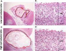
          Contrast T1-weighted magnetic resonance imaging with gadolinium was performed before and after antiparasitic treatment. Eight NCC-infected pigs were treated with praziquantel plus albendazole and euthanized two (n = 4) and five (n = 4) days after treatment; another group of four infected pigs served as untreated controls. For each lesion, gadolinium enhancement intensity (GEI) and cyst volume were measured at baseline and after antiparasitic treatment. Volume and GEI quantification ratios (post/pre-treatment measures) were used to appraise the effect of treatment. Cysts from untreated pigs showed little variations between their basal and post treatment measures. At days 2 and 5 there were significant increases in GEI ratio compared with the untreated group (1.32 and 1.47 vs 1.01, p = 0.021 and p = 0.021). Cyst volume ratios were significantly lower at days 2 and 5 compared with the untreated group (0.60 and 0.22 vs 0.95, p = 0.04 and p = 0.02). Cysts with lower cyst volume ratios showed more marked post-treatment inflammation, loss of vesicular fluid and cyst wall wrinkling.

          Conclusion/Significance

          A significant and drastic reduction of cyst size and increased pericystic enhancement occur in the initial days after antiparasitic treatment as an effect of acute perilesional immune response. These significant changes showed that early anthelmintic efficacy (day two) can be detected using magnetic resonance imaging.

          Author summary

          Neurocysticercosis (NCC) is a frequent parasitic infection of the human brain and the most common cause of adult onset epilepsy in developing countries. Acute inflammatory response in NCC plays an important role in the pathogenesis of symptoms by anthelminitic therapies. The anthelmintic recommended therapy for NCC has drawbacks as the exacerbation of inflammation around degenerating cysts provokes the appearance of symptoms at the first days of treatment. Radiological changes in the appearance of cysts usually are seen after months of therapy. To evaluate if significant radiological changes (enhancement and size) occur in the first days of therapy, we used a porcine NCC model and magnetic resonance imaging (MRI) with contrast solution. The major radiological changes observed after treatment with albendazole and praziquantel were an increase in enhancement and the significant reduction in cyst size by day 2 and more evident on day 5. Cysts with greater changes also experienced exacerbated inflammation, loss of vesicular fluid and wrinkling of the cyst wall. These results show an early therapeutic effect and the possible utility of repeat MRI imaging within a few days after starting treatment. Finally, these findings contribute to our understanding of the treatment induced early exacerbation of symptoms.

          Related collections

          Most cited references36

          • Record: found
          • Abstract: found
          • Article: not found

          Clinical symptoms, diagnosis, and treatment of neurocysticercosis.

          The infection of the nervous system by the cystic larvae of Taenia solium (neurocysticercosis) is a frequent cause of seizure disorders. Neurocysticercosis is endemic or presumed to be endemic in many low-income countries. The lifecycle of the worm and the clinical manifestations of neurocysticercosis are well established, and CT and MRI have substantially improved knowledge of the disease course. Improvements in immunodiagnosis have further advanced comprehension of the pathophysiology of this disease. This knowledge has led to individualised treatment approaches that account for the involvement of parenchymal or extraparenchymal spaces, the number and form of parasites, and the extent of degeneration and associated inflammation. Clinical investigations are focused on development of effective treatments and reduction of side-effects induced by treatment, such as seizures, hydrocephalus, infarcts, and neuroinjury.
            Bookmark
            • Record: found
            • Abstract: found
            • Article: not found

            Diagnosis and treatment of neurocysticercosis.

            Neurocysticercosis is a parasitic disease caused by the larval (cystic) form of the pork cestode tapeworm, Taenia solium, and is a major cause of acquired seizures and epilepsy worldwide. Development of sensitive and specific diagnostic methods, particularly CT and MRI, has revolutionized our knowledge of the burden of cysticercosis infection and disease, and has led to the development of effective antihelminthic treatments for neurocysticercosis. The importance of calcified granulomas with perilesional edema as foci of seizures and epilepsy in populations where neurocysticercosis is endemic is newly recognized, and indicates that treatment with anti-inflammatory agents could have a role in controlling or preventing epilepsy in these patients. Importantly, neurocysticercosis is one of the few diseases that could potentially be controlled or eliminated-an accomplishment that would prevent millions of cases of epilepsy. This Review examines the rationale for treatment of neurocysticercosis and highlights the essential role of inflammation in the pathogenesis of disease, the exacerbation of symptoms that occurs as a result of antihelminthic treatment, and the limitations of current antihelminthic and anti-inflammatory treatments.
              Bookmark
              • Record: found
              • Abstract: found
              • Article: not found

              A trial of antiparasitic treatment to reduce the rate of seizures due to cerebral cysticercosis.

              Neurocysticercosis is the main cause of adult-onset seizures in the developing world. Whether therapy with antiparasitic agents results in improved seizure control has been questioned because of the lack of adequate, controlled studies. We conducted a double-blind, placebo-controlled trial in which 120 patients who had living cysticerci in the brain and seizures treated with antiepileptic drugs were randomly assigned to receive either 800 mg of albendazole per day and 6 mg of dexamethasone per day for 10 days (60 patients) or two placebos (60 patients). The patients were followed for 30 months or until they had been seizure-free for 6 months after the doses of the antiepileptic drugs had been tapered. The efficacy of treatment was measured as the decrease in the number of seizures after treatment. In the albendazole group, there was a 46 percent reduction in the number of seizures (95 percent confidence interval, -74 to 83 percent) during months 2 to 30 after treatment. This reduction, which was not statistically significant, was composed of a nonsignificant reduction of 41 percent in the number of partial seizures (95 percent confidence interval, -124 to 84 percent) and a significant 67 percent reduction in the number of seizures with generalization (95 percent confidence interval, 20 to 86 percent). Most of the difference in the number of partial seizures was attributable to a few patients who had many seizures during follow-up. The proportions of patients who had partial seizures during follow-up were similar in the two groups (19 of 57 in the albendazole group and 16 of 59 in the placebo group), but the patients in the placebo group had a greater tendency to have seizures with generalization (22 of 59, vs. 13 of 57 in the albendazole group; risk ratio, 1.63; 95 percent confidence interval, 0.91 to 2.92). More of the intracranial cystic lesions resolved in the albendazole group than in the placebo group. With the sole exception of abdominal pain, side effects did not differ significantly between the two groups. In patients with seizures due to viable parenchymal cysts, antiparasitic therapy decreases the burden of parasites and is safe and effective, at least in reducing the number of seizures with generalization. Copyright 2004 Massachusetts Medical Society
                Bookmark

                Author and article information

                Contributors
                Role: Editor
                Journal
                PLoS Negl Trop Dis
                PLoS Negl Trop Dis
                plos
                plosntds
                PLoS Neglected Tropical Diseases
                Public Library of Science (San Francisco, CA USA )
                1935-2727
                1935-2735
                2 June 2017
                June 2017
                : 11
                : 6
                : e0005624
                Affiliations
                [1 ]Laboratorio de Inmunopatología en Neurocisticercosis, Facultad de Ciencias y Filosofía, Universidad Peruana Cayetano Heredia, Lima, Peru
                [2 ]Facultad de Medicina Humana, Universidad Nacional Mayor de San Marcos, Lima, Peru
                [3 ]Unidad de Cisticercosis, Instituto Nacional de Ciencias Neurológicas, Lima, Peru
                [4 ]Facultad de Medicina Veterinaria, Universidad Nacional Mayor de San Marcos, Lima, Peru
                [5 ]Facultad de Medicina Veterinaria y Zootecnia, Universidad Peruana Cayetano Heredia, Lima, Peru
                [6 ]Facultad de Medicina, Universidad Peruana Cayetano Heredia, Lima, Peru
                [7 ]Departamento de Diagnóstico por imágenes, Instituto Nacional de Ciencias Neurológicas, Lima, Peru
                [8 ]Departamento de Ciencias Celulares y Moleculares, Facultad de Ciencias y Filosofía, Universidad Peruana Cayetano Heredia, Lima, Peru
                [9 ]Laboratory of Parasitic Diseases, National Institute of Allergy and Infectious Diseases, National Institutes of Health, Bethesda, Maryland, United States of America
                Michigan State University, UNITED STATES
                Author notes

                The authors have declared that no competing interests exist.

                • Conceptualization: JAB HHG TEN CC.

                • Data curation: CC HHG JAB.

                • Formal analysis: CC HHG TEN JAB.

                • Funding acquisition: HHG JAB TEN SM CGG.

                • Investigation: JAB HHG CC CGG JM DS JCh GA JCa AVC.

                • Methodology: HHG TEN JAB CC JCa AVC CGG.

                • Project administration: HHG JAB TEN SM CGG.

                • Resources: HHG JAB CGG AEG TEN SM.

                • Software: CC HHG TEN JAB.

                • Supervision: HHG JAB TEN SM CGG CC.

                • Validation: CC HHG TEN JAB.

                • Visualization: CC HHG TEN SM CGG JAB.

                • Writing – original draft: CC HHG TEN SM CGG JAB.

                • Writing – review & editing: CC HHG TEN SM CGG JAB.

                ¶ Membership of the Cysticercosis Working Group in Peru is provided in the Acknowledgments.

                Author information
                http://orcid.org/0000-0002-3525-8445
                Article
                PNTD-D-16-02044
                10.1371/journal.pntd.0005624
                5470720
                28575043
                eb30cb7e-994a-49b4-9057-59a3b0b7b29b

                This is an open access article, free of all copyright, and may be freely reproduced, distributed, transmitted, modified, built upon, or otherwise used by anyone for any lawful purpose. The work is made available under the Creative Commons CC0 public domain dedication.

                History
                : 10 November 2016
                : 4 May 2017
                Page count
                Figures: 3, Tables: 3, Pages: 14
                Funding
                This work was supported in part by the Intramural Research Program of the National Institute of Allergy and Infectious Diseases, and by FIC-NIH training grant TW001140, NIH 1R01AI116456, and Beca Anual de Medicina “Francisco Tejada y Semíramis Reátegui” 2014 Universidad Peruana Cayetano Heredia. HHG is supported by a Wellcome Trust Senior International Research Fellowship in Public Health and Tropical Medicine. The funders had no role in study design, data collection and analysis, decision to publish, or preparation of the manuscript.
                Categories
                Research Article
                Biology and Life Sciences
                Organisms
                Animals
                Vertebrates
                Amniotes
                Mammals
                Swine
                Biology and Life Sciences
                Immunology
                Immune Response
                Inflammation
                Medicine and Health Sciences
                Immunology
                Immune Response
                Inflammation
                Medicine and Health Sciences
                Diagnostic Medicine
                Signs and Symptoms
                Inflammation
                Medicine and Health Sciences
                Pathology and Laboratory Medicine
                Signs and Symptoms
                Inflammation
                Medicine and Health Sciences
                Diagnostic Medicine
                Diagnostic Radiology
                Magnetic Resonance Imaging
                Research and Analysis Methods
                Imaging Techniques
                Diagnostic Radiology
                Magnetic Resonance Imaging
                Medicine and Health Sciences
                Radiology and Imaging
                Diagnostic Radiology
                Magnetic Resonance Imaging
                Biology and Life Sciences
                Immunology
                Vaccination and Immunization
                Antiparasitic Therapy
                Medicine and Health Sciences
                Immunology
                Vaccination and Immunization
                Antiparasitic Therapy
                Medicine and Health Sciences
                Public and Occupational Health
                Preventive Medicine
                Vaccination and Immunization
                Antiparasitic Therapy
                Medicine and Health Sciences
                Radiology and Imaging
                Research and Analysis Methods
                Experimental Organism Systems
                Animal Models
                Pig Models
                Physical Sciences
                Chemistry
                Chemical Elements
                Gadolinium
                Medicine and Health Sciences
                Pharmaceutics
                Drug Therapy
                Custom metadata
                vor-update-to-uncorrected-proof
                2017-06-14
                All relevant data are within the paper and its Supporting Information files.

                Infectious disease & Microbiology
                Infectious disease & Microbiology

                Comments

                Comment on this article