0
views
0
recommends
+1 Recommend
0 collections
    0
    shares
      • Record: found
      • Abstract: found
      • Article: found
      Is Open Access

      β-hydroxybutyrate accumulates in the rat heart during low-flow ischaemia with implications for functional recovery

      research-article

      Read this article at

      Bookmark
          There is no author summary for this article yet. Authors can add summaries to their articles on ScienceOpen to make them more accessible to a non-specialist audience.

          Abstract

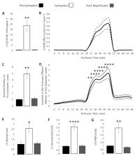
          Extrahepatic tissues which oxidise ketone bodies also have the capacity to accumulate them under particular conditions. We hypothesised that acetyl-coenzyme A (acetyl-CoA) accumulation and altered redox status during low-flow ischaemia would support ketone body production in the heart. Combining a Langendorff heart model of low-flow ischaemia/reperfusion with liquid chromatography coupled tandem mass spectrometry (LC-MS/MS), we show that β-hydroxybutyrate (β-OHB) accumulated in the ischaemic heart to 23.9 nmol/gww and was secreted into the coronary effluent. Sodium oxamate, a lactate dehydrogenase (LDH) inhibitor, increased ischaemic β-OHB levels 5.3-fold and slowed contractile recovery. Inhibition of β-hydroxy-β-methylglutaryl (HMG)-CoA synthase (HMGCS2) with hymeglusin lowered ischaemic β-OHB accumulation by 40%, despite increased flux through succinyl-CoA-3-oxaloacid CoA transferase (SCOT), resulting in greater contractile recovery. Hymeglusin also protected cardiac mitochondrial respiratory capacity during ischaemia/reperfusion. In conclusion, net ketone generation occurs in the heart under conditions of low-flow ischaemia. The process is driven by flux through both HMGCS2 and SCOT, and impacts on cardiac functional recovery from ischaemia/reperfusion.

          Related collections

          Most cited references35

          • Record: found
          • Abstract: found
          • Article: found

          Ischaemic accumulation of succinate controls reperfusion injury through mitochondrial ROS

          Ischaemia-reperfusion (IR) injury occurs when blood supply to an organ is disrupted and then restored, and underlies many disorders, notably heart attack and stroke. While reperfusion of ischaemic tissue is essential for survival, it also initiates oxidative damage, cell death, and aberrant immune responses through generation of mitochondrial reactive oxygen species (ROS) 1-5 . Although mitochondrial ROS production in IR is established, it has generally been considered a non-specific response to reperfusion 1,3 . Here, we developed a comparative in vivo metabolomic analysis and unexpectedly identified widely conserved metabolic pathways responsible for mitochondrial ROS production during IR. We showed that selective accumulation of the citric acid cycle (CAC) intermediate succinate is a universal metabolic signature of ischaemia in a range of tissues and is responsible for mitochondrial ROS production during reperfusion. Ischaemic succinate accumulation arises from reversal of succinate dehydrogenase (SDH), which in turn is driven by fumarate overflow from purine nucleotide breakdown and partial reversal of the malate/aspartate shuttle. Upon reperfusion, the accumulated succinate is rapidly re-oxidised by SDH, driving extensive ROS generation by reverse electron transport (RET) at mitochondrial complex I. Decreasing ischaemic succinate accumulation by pharmacological inhibition is sufficient to ameliorate in vivo IR injury in murine models of heart attack and stroke. Thus, we have identified a conserved metabolic response of tissues to ischaemia and reperfusion that unifies many hitherto unconnected aspects of IR injury. Furthermore, these findings reveal a novel pathway for metabolic control of ROS production in vivo, while demonstrating that inhibition of ischaemic succinate accumulation and its oxidation upon subsequent reperfusion is a potential therapeutic target to decrease IR injury in a range of pathologies.
            Bookmark
            • Record: found
            • Abstract: found
            • Article: not found

            Multi-dimensional Roles of Ketone Bodies in Fuel Metabolism, Signaling, and Therapeutics

            Ketone body metabolism is a central node in physiological homeostasis. In this review, we discuss how ketones serve discrete fine-tuning metabolic roles that optimize organ and organism performance in varying nutrient states and protect from inflammation and injury in multiple organ systems. Traditionally viewed as metabolic substrates enlisted only in carbohydrate restriction, observations underscore the importance of ketone bodies as vital metabolic and signaling mediators when carbohydrates are abundant. Complementing a repertoire of known therapeutic options for diseases of the nervous system, prospective roles for ketone bodies in cancer have arisen, as have intriguing protective roles in heart and liver, opening therapeutic options in obesity-related and cardiovascular disease. Controversies in ketone metabolism and signaling are discussed to reconcile classical dogma with contemporary observations.
              Bookmark
              • Record: found
              • Abstract: found
              • Article: not found

              High-resolution respirometry: OXPHOS protocols for human cells and permeabilized fibers from small biopsies of human muscle.

              Protocols for high-resolution respirometry (HRR) of intact cells, permeabilized cells, and permeabilized muscle fibers offer sensitive diagnostic tests of integrated mitochondrial function using standard cell culture techniques and small needle biopsies of muscle. Multiple substrate-uncoupler-inhibitor titration (SUIT) protocols for analysis of oxidative phosphorylation improve our understanding of mitochondrial respiratory control and the pathophysiology of mitochondrial diseases. Respiratory states are defined in functional terms to account for the network of metabolic interactions in complex SUIT protocols with stepwise modulation of coupling and substrate control. A regulated degree of intrinsic uncoupling is a hallmark of oxidative phosphorylation, whereas pathological and toxicological dyscoupling is evaluated as a mitochondrial defect. The noncoupled state of maximum respiration is experimentally induced by titration of established uncouplers (FCCP, DNP) to collapse the proton gradient across the mitochondrial inner membrane and measure the capacity of the electron transfer system (ETS, open-circuit operation of respiration). Intrinsic uncoupling and dyscoupling are evaluated as the flux control ratio between nonphosphorylating LEAK respiration (electron flow coupled to proton pumping to compensate for proton leaks) and ETS capacity. If OXPHOS capacity (maximally ADP-stimulated oxygen flux) is less than ETS capacity, the phosphorylation system contributes to flux control. Physiological Complex I + II substrate combinations are required to reconstitute TCA cycle function. This supports maximum ETS and OXPHOS capacities, due to the additive effect of multiple electron supply pathways converging at the Q-junction. Substrate control with electron entry separately through Complex I (pyruvate + malate or glutamate + malate) or Complex II (succinate + rotenone) restricts ETS capacity and artificially enhances flux control upstream of the Q-cycle, providing diagnostic information on specific branches of the ETS. Oxygen levels are maintained above air saturation in protocols with permeabilized muscle fibers to avoid experimental oxygen limitation of respiration. Standardized two-point calibration of the polarographic oxygen sensor (static sensor calibration), calibration of the sensor response time (dynamic sensor calibration), and evaluation of instrumental background oxygen flux (systemic flux compensation) provide the unique experimental basis for high accuracy of quantitative results and quality control in HRR.
                Bookmark

                Author and article information

                Contributors
                Role: Senior Editor
                Role: Reviewing Editor
                Journal
                eLife
                Elife
                eLife
                eLife
                eLife Sciences Publications, Ltd
                2050-084X
                07 September 2021
                2021
                : 10
                : e71270
                Affiliations
                [1 ] Department of Physiology, Development and Neuroscience, University of Cambridge London United Kingdom
                [2 ] Department of Biochemistry and Cambridge Systems Biology Centre, University of Cambridge London United Kingdom
                [3 ] Section of Biomolecular Medicine, Systems Medicine, Department of Metabolism, Digestion and Reproduction, Imperial College London London United Kingdom
                University of Zurich Switzerland
                Flinders Medical Centre Australia
                Flinders Medical Centre Australia
                University ofTexas Health Science Center at Houston United States
                Author notes
                [†]

                Research and Early Development, Cardiovascular, Renal and Metabolic Diseases, BioPharmaceuticals R&D, AstraZeneca Ltd, Cambridge, United Kingdom.

                Author information
                https://orcid.org/0000-0001-7760-613X
                Article
                71270
                10.7554/eLife.71270
                8423437
                34491199
                ebdeb3bf-5ddd-4428-8456-d2229cdeb4d4
                © 2021, Lindsay et al

                This article is distributed under the terms of the Creative Commons Attribution License, which permits unrestricted use and redistribution provided that the original author and source are credited.

                History
                : 15 June 2021
                : 23 August 2021
                Funding
                Funded by: FundRef http://dx.doi.org/10.13039/501100000274, British Heart Foundation;
                Award ID: FS/14/59/31282
                Award Recipient :
                Funded by: FundRef http://dx.doi.org/10.13039/501100000690, Research Councils UK;
                Award ID: EP/E500552/1
                Award Recipient :
                The funders had no role in study design, data collection and interpretation, or the decision to submit the work for publication.
                Categories
                Research Article
                Biochemistry and Chemical Biology
                Cell Biology
                Custom metadata
                β-Hydroxybutyrate accumulates in the perfused rat heart during ischaemia, driven by flux through both HMGCS and SCOT, with implications for functional recovery.

                Life sciences
                heart,langendorff,cardiomyocyte,ischaemia,rat
                Life sciences
                heart, langendorff, cardiomyocyte, ischaemia, rat

                Comments

                Comment on this article