9
views
0
recommends
+1 Recommend
0 collections
    0
    shares
      • Record: found
      • Abstract: found
      • Article: found
      Is Open Access

      The characteristics of Laennec's capsule around the hepatic veins: A histological study based on 71 liver surgical specimens

      research-article

      Read this article at

      Bookmark
          There is no author summary for this article yet. Authors can add summaries to their articles on ScienceOpen to make them more accessible to a non-specialist audience.

          Abstract

          Background

          Laennec's capsule is a fibrous membrane attached to the surface of the liver, which is independent of the hepatic veins. However, the presence of Laennec's capsule surrounding the peripheral hepatic veins is controversial. This study aims to describe the characteristic of Laennec's capsule around the hepatic veins at all levels.

          Methods

          Seventy‐one hepatic surgical specimens were collected along the cross and longitudinal sections of the hepatic vein. Tissue sections of 3–4 mm were cut and stained with hematoxylin and eosin (H&E), resorcinol‐fuchsin (R&F), and Victoria blue (V&B). Elastic fibers were observed around the hepatic veins. They were measured using K‐Viewer software.

          Results

          Morphologically, we observed a thin, dense fibrous layer (so‐called Laennec's capsule) around the hepatic veins at all levels, which was different from the thick elastic fibers of the hepatic vein wall. Therefore, there was a potential gap between Laennec's capsule and the hepatic veins. Laennec's capsule was visualized significantly better with R&F and V&B staining compared to H&E staining. The thickness of Laennec's capsule around the main, first, and secondary branches of the hepatic vein were 79.86 ± 24.20 μm, 48.41 ± 18.25 μm, and 23.56 ± 10.03 μm in the R&F staining, and 80.15 ± 21.85 μm, 49.46 ± 17.52 μm, and 25.05 ± 11.03 μm in the V&B staining, respectively. They were significantly different from each other ( P < .001).

          Conclusion

          The hepatic veins were surrounded by Laennec's capsule at all levels, including the peripheral hepatic veins. However, it is thinner along the vein branches. The gap between the Laennec's capsule and hepatic veins shows potential supplemental value for liver surgery.

          Abstract

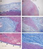
          Histological findings of Laennec's capsule in the liver tissues of the hepatic veins after hematoxylin and eosin (H&E) (A, D, G and J), resorcinol‐fuchsin (R&F) (B, E, H, K and M), and Victoria blue (V&B) (C, F, I, L and N) staining, such as the main hepatic vein (A‐C), the first branch of hepatic vein (D‐F), the secondary branch of hepatic vein (G‐I), the inferior vena cava (J‐L) and the umbilical veins as the control group (M, N). The outer layer of the hepatic vein is composed of thick and loose smooth muscle fibers (blue triangle). The same is true for the inferior vena cava and the umbilical vein. Laennec's capsule (arrows and brackets) is a dense and smooth thin membrane structure, which does not contain thick elastic fibers

          Related collections

          Most cited references13

          • Record: found
          • Abstract: found
          • Article: found
          Is Open Access

          Systematic extrahepatic Glissonean pedicle isolation for anatomical liver resection based on Laennec's capsule: proposal of a novel comprehensive surgical anatomy of the liver

          Abstract Anatomical liver resection with the Glissonean pedicle isolation is widely approved as an essential procedure for safety and curability. Especially, the extrahepatic Glissonean pedicle isolation without parenchymal destruction should be an ideal procedure. However, the surgical technique has not been standardized due to a lack of anatomical understanding. Herein, we proposed a novel comprehensive surgical anatomy of the liver based on Laennec's capsule that would give a theoretical background to the extrahepatic Glissonean pedicle isolation. Laennec's capsule is the proper membrane that covers not only the entire surface of the liver including the bare area but also the intrahepatic parenchyma surrounding the Glissonean pedicles. Consequently, there exists a gap between the Glissonean pedicle and Laennec's capsule that could be reached extrahepatically and allows us to isolate the extrahepatic Glissonean pedicle without parenchymal destruction systematically. For standardization, it is essential to approach the “six gates” indicated by the “four anatomical landmarks”: the Arantius plate, the umbilical plate, the cystic plate and the Glissonean pedicle of the caudate process (G1c). This novel anatomy would contribute to standardize the surgical techniques of the systematic extrahepatic Glissonean pedicle isolation for anatomical liver resection including laparoscopic or robotic liver resection and to bring innovative changes in hepatobiliary surgery for spreading safe and curable liver resection.
            Bookmark
            • Record: found
            • Abstract: found
            • Article: not found

            Comparison of anatomic and non-anatomic hepatic resection for hepatocellular carcinoma

            The aim of the present study was to compare the prognostic impact of anatomic resection (AR) versus non-anatomic resection (NAR) on patient survival after resection of a single hepatocellular carcinoma (HCC).
              Bookmark
              • Record: found
              • Abstract: found
              • Article: not found

              Connective tissue configuration in the human liver hilar region with special reference to the liver capsule and vascular sheath.

              We carried out this study to examine the validity of the accepted dogma that: (1) the human liver capsule does not extend along the fissures for the hepatic veins; (2) the hilar vasculobiliary sheath does not connect to the liver capsule; and (3) the hilar plate is a thickening of the vasculobiliary sheath. Using cadaveric specimens, we identified composite fibers and other structures in the sheath and capsule histologically. The liver capsule, Glisson's sheath, and the sheath for the hepatic vein tributaries were characterized by a high content of thin, wavy elastic fibers. However, the hilar vasculobiliary sheath of the thick vessels and ducts did not contain elastic fibers. Along the roof of the hilar region, vaginal ductuli were identified as a chain of cross-sectional bile ducts with a relatively thick wall, because of their tortuous course with abundant small pouches budding from the surface. The ductuli were separated from the liver capsule by abundant lymphatic vessels. The sheath for hepatic veins and Glisson's sheath appear to connect to, and be continuous with, the liver capsule. During surgery and dissection, it should be borne in mind that the hilar plate is likely to be artificially developed when, without intention, surgeon bundle collagenous fibers with vaginal ductuli forming a core.
                Bookmark

                Author and article information

                Contributors
                kinghaos@126.com
                Journal
                Ann Gastroenterol Surg
                Ann Gastroenterol Surg
                10.1002/(ISSN)2475-0328
                AGS3
                Annals of Gastroenterological Surgery
                John Wiley and Sons Inc. (Hoboken )
                2475-0328
                27 September 2022
                March 2023
                : 7
                : 2 ( doiID: 10.1002/ags3.v7.2 )
                : 287-294
                Affiliations
                [ 1 ] Department of General Surgery, Guangdong Provincial People's Hospital Guangdong Academy of Medical Sciences Guangzhou China
                [ 2 ] Guangdong Cardiovascular Institute Guangzhou China
                [ 3 ] Shantou University Medical College Shantou China
                [ 4 ] Department of Pathology, Guangdong Provincial People's Hospital Guangdong Academy of Medical Sciences Guangzhou China
                Author notes
                [*] [* ] Correspondence

                Haosheng Jin, Department of General Surgery, Guangdong Provincial People's Hospital, Guangdong Academy of Medical Sciences, Guangzhou, China, No. 106, Zhongshan 2nd Road, Guangzhou, CN 510080, China.

                Email: kinghaos@ 123456126.com

                Author information
                https://orcid.org/0000-0002-3681-3930
                Article
                AGS312618 AGS-2022-0192.R1
                10.1002/ags3.12618
                10043764
                ec29f8e6-4681-42c2-893f-7b635046e698
                © 2022 The Authors. Annals of Gastroenterological Surgery published by John Wiley & Sons Australia, Ltd on behalf of The Japanese Society of Gastroenterological Surgery.

                This is an open access article under the terms of the http://creativecommons.org/licenses/by-nc/4.0/ License, which permits use, distribution and reproduction in any medium, provided the original work is properly cited and is not used for commercial purposes.

                History
                : 25 May 2022
                : 19 August 2022
                Page count
                Figures: 4, Tables: 2, Pages: 8, Words: 4319
                Funding
                Funded by: Natural Science Foundation of Guangdong Province , doi 10.13039/501100003453;
                Award ID: 2021A1515011066
                Categories
                Original Article
                Original Articles
                Custom metadata
                2.0
                March 2023
                Converter:WILEY_ML3GV2_TO_JATSPMC version:6.2.6 mode:remove_FC converted:28.03.2023

                elastic fibers,hepatic vein,laennec's capsule,live surgery,liver

                Comments

                Comment on this article