45
views
0
recommends
+1 Recommend
0 collections
    0
    shares
      • Record: found
      • Abstract: found
      • Article: not found

      Targeting cellular senescence prevents age-related bone loss in mice

      research-article

      Read this article at

      ScienceOpenPublisherPMC
      Bookmark
          There is no author summary for this article yet. Authors can add summaries to their articles on ScienceOpen to make them more accessible to a non-specialist audience.

          Abstract

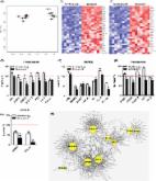
          Aging is associated with increased cellular senescence, which is hypothesized to drive the eventual development of multiple co-morbidities 1 . Here, we investigate a role for senescent cells in age-related bone loss by multiple approaches. In particular, we used either genetic ( i.e., the INK-ATTAC “suicide” transgene encoding an inducible caspase 8 expressed specifically in senescent cells 24 ) or pharmacological ( i.e., “senolytic” compounds 5, 6 ) means to eliminate senescent cells. We also inhibited the production of the pro-inflammatory secretome of senescent cells using a JAK inhibitor (JAKi) 3, 7 . In old (20–22-months) mice with established bone loss, activation of the INK-ATTAC caspase 8 in senescent cells or treatment with senolytics or the JAKi for 2–4 months resulted in higher bone mass and strength and better bone microarchitecture compared to vehicle-treated mice. The beneficial effects of targeting senescent cells were due to lower bone resorption with either maintained (trabecular bone) or higher (cortical bone) bone formation as compared to vehicle-treated mice. In vitro studies demonstrated that senescent cell-conditioned medium impaired osteoblast mineralization and enhanced osteoclast progenitor survival, leading to increased osteoclastogenesis. Collectively, these data establish a causal role for senescent cells in bone loss with aging and demonstrate that targeting these cells has both anti-resorptive and anabolic effects on bone. As eliminating senescent cells and/or inhibiting their pro-inflammatory secretome also improves cardiovascular function 4 , enhances insulin sensitivity 3 , and reduces frailty 7 , targeting this fundamental mechanism to prevent age-related bone loss suggests a novel treatment strategy not only for osteoporosis but also for multiple age-related co-morbidities.

          Related collections

          Most cited references27

          • Record: found
          • Abstract: found
          • Article: not found

          An improved technique for determining hardness and elastic modulus using load and displacement sensing indentation experiments

          The indentation load-displacement behavior of six materials tested with a Berkovich indenter has been carefully documented to establish an improved method for determining hardness and elastic modulus from indentation load-displacement data. The materials included fused silica, soda–lime glass, and single crystals of aluminum, tungsten, quartz, and sapphire. It is shown that the load–displacement curves during unloading in these materials are not linear, even in the initial stages, thereby suggesting that the flat punch approximation used so often in the analysis of unloading data is not entirely adequate. An analysis technique is presented that accounts for the curvature in the unloading data and provides a physically justifiable procedure for determining the depth which should be used in conjunction with the indenter shape function to establish the contact area at peak load. The hardnesses and elastic moduli of the six materials are computed using the analysis procedure and compared with values determined by independent means to assess the accuracy of the method. The results show that with good technique, moduli can be measured to within 5%.
            Bookmark
            • Record: found
            • Abstract: found
            • Article: found
            Is Open Access

            The Achilles’ heel of senescent cells: from transcriptome to senolytic drugs

            The healthspan of mice is enhanced by killing senescent cells using a transgenic suicide gene. Achieving the same using small molecules would have a tremendous impact on quality of life and the burden of age-related chronic diseases. Here, we describe the rationale for identification and validation of a new class of drugs termed senolytics, which selectively kill senescent cells. By transcript analysis, we discovered increased expression of pro-survival networks in senescent cells, consistent with their established resistance to apoptosis. Using siRNA to silence expression of key nodes of this network, including ephrins (EFNB1 or 3), PI3Kδ, p21, BCL-xL, or plasminogen-activated inhibitor-2, killed senescent cells, but not proliferating or quiescent, differentiated cells. Drugs targeting these same factors selectively killed senescent cells. Dasatinib eliminated senescent human fat cell progenitors, while quercetin was more effective against senescent human endothelial cells and mouse BM-MSCs. The combination of dasatinib and quercetin was effective in eliminating senescent MEFs. In vivo, this combination reduced senescent cell burden in chronologically aged, radiation-exposed, and progeroid Ercc1 −/Δ mice. In old mice, cardiac function and carotid vascular reactivity were improved 5 days after a single dose. Following irradiation of one limb in mice, a single dose led to improved exercise capacity for at least 7 months following drug treatment. Periodic drug administration extended healthspan in Ercc1 −/Δ mice, delaying age-related symptoms and pathology, osteoporosis, and loss of intervertebral disk proteoglycans. These results demonstrate the feasibility of selectively ablating senescent cells and the efficacy of senolytics for alleviating symptoms of frailty and extending healthspan.
              Bookmark
              • Record: found
              • Abstract: found
              • Article: not found

              The recent prevalence of osteoporosis and low bone mass in the United States based on bone mineral density at the femoral neck or lumbar spine.

              The goal of our study was to estimate the prevalence of osteoporosis and low bone mass based on bone mineral density (BMD) at the femoral neck and the lumbar spine in adults 50 years and older in the United States (US). We applied prevalence estimates of osteoporosis or low bone mass at the femoral neck or lumbar spine (adjusted by age, sex, and race/ethnicity to the 2010 Census) for the noninstitutionalized population aged 50 years and older from the National Health and Nutrition Examination Survey 2005-2010 to 2010 US Census population counts to determine the total number of older US residents with osteoporosis and low bone mass. There were more than 99 million adults aged 50 years and older in the US in 2010. Based on an overall 10.3% prevalence of osteoporosis, we estimated that in 2010, 10.2 million older adults had osteoporosis. The overall low bone mass prevalence was 43.9%, from which we estimated that 43.4 million older adults had low bone mass. We estimated that 7.7 million non-Hispanic white, 0.5 million non-Hispanic black, and 0.6 million Mexican American adults had osteoporosis, and another 33.8, 2.9, and 2.0 million had low bone mass, respectively. When combined, osteoporosis and low bone mass at the femoral neck or lumbar spine affected an estimated 53.6 million older US adults in 2010. Although most of the individuals with osteoporosis or low bone mass were non-Hispanic white women, a substantial number of men and women from other racial/ethnic groups also had osteoporotic BMD or low bone mass. © 2014 American Society for Bone and Mineral Research.
                Bookmark

                Author and article information

                Journal
                9502015
                8791
                Nat Med
                Nat. Med.
                Nature medicine
                1078-8956
                1546-170X
                17 July 2017
                21 August 2017
                September 2017
                21 February 2018
                : 23
                : 9
                : 1072-1079
                Affiliations
                Robert and Arlene Kogod Center on Aging and Division of Endocrinology, Mayo Clinic College of Medicine, Rochester, MN, 55905
                Author notes
                [* ]Co-corresponding authors: Sundeep Khosla, M.D., College of Medicine, Mayo Clinic, 200 First Street SW, Rochester, Minnesota 55905; Tel: 507-255-6663; khosla.sundeep@ 123456mayo.edu . James L. Kirkland, M.D., Ph.D., College of Medicine, Mayo Clinic, 200 First Street SW, Rochester, Minnesota 55905; Tel: 507-266-9151; kirkland.james@ 123456mayo.edu
                [†]

                These authors contributed equally to this work.

                Article
                NIHMS892924
                10.1038/nm.4385
                5657592
                28825716
                ee1a4eb9-af9a-40c9-874f-e1a591f43315

                Users may view, print, copy, and download text and data-mine the content in such documents, for the purposes of academic research, subject always to the full Conditions of use: http://www.nature.com/authors/editorial_policies/license.html#terms

                History
                Categories
                Article

                Medicine
                aging,osteocyte,osteoclast,disease prevention,drug therapy
                Medicine
                aging, osteocyte, osteoclast, disease prevention, drug therapy

                Comments

                Comment on this article