1
views
0
recommends
+1 Recommend
0 collections
    0
    shares
      • Record: found
      • Abstract: found
      • Article: found
      Is Open Access

      Not All Hepatocellular Carcinoma Patients with Microvascular Invasion After R0 Resection Could Be Benefited from Prophylactic Transarterial Chemoembolization: A Propensity Score Matching Study

      research-article

      Read this article at

      Bookmark
          There is no author summary for this article yet. Authors can add summaries to their articles on ScienceOpen to make them more accessible to a non-specialist audience.

          Abstract

          Background

          Prophylactic transarterial chemoembolization (p-TACE) is strongly recommended for hepatocellular carcinoma (HCC) patients with microvascular invasion (MVI), but the potential beneficiaries remain controversial.

          Methods

          Data of HCC patients with MVI who underwent R0 resection between December 2013 and December 2015 were identified through the primary liver cancer big data. Disease-free survival (DFS) and overall survival (OS) were compared between patients who received p-TACE or not using Kaplan–Meier survival curves before and after propensity scoring match (PSM).

          Results

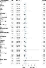
          A total of 695 patients were eligible for this study, including 199 patients (28.6%) receiving p-TACE and 496 patients (71.4%) receiving resection alone. In the crude cohort, median DFS and OS were longer in the p-TACE group than those in the non-TACE group without significant differences (25.0 months vs 24.2 months, P=0.100; 48.0 months vs 46.5 months, P=0.150; respectively), but significant differences were observed both in DFS and OS (both P<0.05) after 1:1 PSM. p-TACE was identified as one of the independent risk factors of both DFS and OS using multivariate analysis in the matched cohort (HR=0.69, 95% CI=0.54–0.88; HR=0.66, 95% CI=0.50–0.88; respectively). Subgroup analysis showed that p-TACE could beneficiate patients if they were male, aged ≥50 years old, had HBV infection, preoperative AFP level ≥400 ng/mL, Child-Pugh grading A, no transfusion, single tumor, tumor diameter ≥5cm, Edmondson–Steiner grading I/II, capsule, or BCLC stage A, CNLC stage Ib, AJCC stage II both in DFS and OS (all P<0.05).

          Conclusion

          With the current data, we concluded that not all HCC patients with MVI would be benefited from p-TACE, and p-TACE could benefit patients with “middle risk” according to the current staging systems.

          Related collections

          Most cited references25

          • Record: found
          • Abstract: found
          • Article: found

          Guidelines for Diagnosis and Treatment of Primary Liver Cancer in China (2017 Edition)

          Background: Hepatocellular carcinoma (HCC) (about 85–90% of primary liver cancer) is particularly prevalent in China because of the high prevalence of chronic hepatitis B infection. HCC is the fourth most common malignancy and the third leading cause of tumor-related deaths in China. It poses a significant threat to the life and health of Chinese people. Summary: This guideline presents official recommendations of the National Health and Family Planning Commission of the People’s Republic of China on the surveillance, diagnosis, staging, and treatment of HCC occurring in China. The guideline was written by more than 50 experts in the field of HCC in China (including liver surgeons, medical oncologists, hepatologists, interventional radiologists, and diagnostic radiologists) on the basis of recent evidence and expert opinions, balance of benefits and harms, cost-benefit strategies, and other clinical considerations. Key Messages: The guideline presents the Chinese staging system, and recommendations regarding patients with HCC in China to ensure optimum patient outcomes.
            Bookmark
            • Record: found
            • Abstract: found
            • Article: not found

            A systematic review of microvascular invasion in hepatocellular carcinoma: diagnostic and prognostic variability.

            Selected patients with hepatocellular carcinoma are candidates to receive potentially curative treatments, such as hepatic resection or liver transplantation, but nevertheless there is a high risk of tumor recurrence. Microvascular invasion is a histological feature of hepatocellular carcinoma related to aggressive biological behavior. We systematically reviewed 20 observational studies that addressed the prognostic impact of microvascular invasion, either after liver transplantation or resection. Outcomes were disease-free survival and overall survival. In liver transplantation, the presence of microvascular invasion shortened disease-free survival at 3 years (relative risk (RR)=3.41 [2.05-5.7]; five studies, n=651) and overall survival both at 3 years (RR=2.41 [1.72-3.37]; five studies, n=1,938) and 5 years (RR=2.29 [1.85-2.83]; six studies, n=2,003). After liver resection, microvascular invasion impacted disease-free survival at 3 and 5 years (RR=1.82 [1.61-2.07] and RR=1.51 [1.29-1.77]; four studies, n=1,501 for both comparisons). However inter/intraobserver variability in reporting and the lack of definition and grading of microvascular invasion has led to great heterogeneity in evaluating this histological feature in hepatocellular carcinoma. Thus, there is an urgent need to clarify this issue, because determining prognosis and response to therapy have become important in the current management of hepatocellular carcinoma. In this systematic review, we summarize the diagnostic and prognostic data concerning microvascular invasion in hepatocellular carcinoma and present a basis for consensus on its definition.
              Bookmark
              • Record: found
              • Abstract: found
              • Article: not found

              Microvascular invasion in patients with hepatocellular carcinoma and its predictable clinicopathological factors.

              Macroscopic vascular invasion is known to be a poor prognostic factor in hepatocellular carcinoma (HCC). The aim of this study was to determine the outcomes and predictive factors after hepatic resection for HCC with microvascular invasion (MVI). One hundred ten patients who underwent curative resection for HCC without macroscopic vascular invasion were included in this retrospective study. The risk factors of these patients for recurrence-free and disease-specific survival were investigated, and the clinicopathological factors predicting the presence of MVI were also determined. Of the 110 resected specimens, 49 (45%) had evidence of MVI. By univariate analysis, MVI was found to be statistically significantly associated with greater tumor size, gross classification, histological grade, and intrahepatic micrometastasis. Gross classification proved to be the only independent predictive factor for MVI by multiple logistic regression analysis. By multivariate analysis, cirrhosis and MVI were identified as independent risk factors for recurrence-free survival. The 5-year recurrence-free survival rates for patients with and without MVI were 20.8% and 52.6%, respectively. By multivariate analysis, the number of tumors, presence of MVI, and intrahepatic micrometastasis were identified as independent predictors of disease-specific survival. The 5-year disease-specific survival rates for patients with and without MVI were 59.3% and 92.0%, respectively. The presence of MVI was the most important risk factor affecting recurrence and survival in HCC patients after curative resection. Furthermore, this study showed that gross classification of HCC can be very helpful in predicting the presence of MVI.
                Bookmark

                Author and article information

                Journal
                Cancer Manag Res
                Cancer Manag Res
                CMAR
                cancmanres
                Cancer Management and Research
                Dove
                1179-1322
                22 May 2020
                2020
                : 12
                : 3815-3825
                Affiliations
                [1 ]Department of Radiation Oncology, Mengchao Hepatobiliary Hospital of Fujian Medical University , Fuzhou, People’s Republic of China
                [2 ]Department of Hepatopancreatobiliary Surgery, Mengchao Hepatobiliary Hospital of Fujian Medical University , Fuzhou, People’s Republic of China
                [3 ]Department of Oncology, Shengli Clinical Medical College of Fujian Medical University, Fujian Provincial Hospital , Fuzhou, People’s Republic of China
                [4 ]Department of Pediatric Surgery, Huai’an Women and Children’s Hospital , Huaian, People’s Republic of China
                [5 ]Department of General Surgery, 900th Hospital of PLA , Fuzhou, People’s Republic of China
                [6 ]The United Innovation of Mengchao Hepatobiliary Technology Key of Fujian Province, Mengchao Hepatobiliary Hospital of Fujian Medical University , Fuzhou, People’s Republic of China
                Author notes
                Correspondence: Jingfeng Liu Tel +86 139 0502 9580Fax +86 591 8370 2529 Email drjingfeng@126.com
                [*]

                These authors contributed equally to this work

                Author information
                http://orcid.org/0000-0001-8975-0489
                http://orcid.org/0000-0001-8886-4054
                http://orcid.org/0000-0003-3499-5678
                Article
                251605
                10.2147/CMAR.S251605
                7250178
                32547217
                f1fec894-58d0-42a4-8060-4e2d1e6acb4e
                © 2020 Wang et al.

                This work is published and licensed by Dove Medical Press Limited. The full terms of this license are available at https://www.dovepress.com/terms.php and incorporate the Creative Commons Attribution – Non Commercial (unported, v3.0) License ( http://creativecommons.org/licenses/by-nc/3.0/). By accessing the work you hereby accept the Terms. Non-commercial uses of the work are permitted without any further permission from Dove Medical Press Limited, provided the work is properly attributed. For permission for commercial use of this work, please see paragraphs 4.2 and 5 of our Terms ( https://www.dovepress.com/terms.php).

                History
                : 27 February 2020
                : 06 May 2020
                Page count
                Figures: 4, Tables: 2, References: 38, Pages: 11
                Funding
                Funded by: Fujian provincial medical center of hepatobiliary, Key Clinical Specialty Discipline Construction Program of Fuzhou
                Award ID: 201912002
                Funded by: Science and Technology project of Fuzhou
                Award ID: 2019-SZ-47
                Funded by: Startup Fund for scientific research, Fujian Medical University
                Award ID: 2018QH1195
                This study was supported by Fujian provincial medical center of hepatobiliary, Key Clinical Specialty Discipline Construction Program of Fuzhou (201912002), Science and Technology project of Fuzhou (Grant number: 2019-SZ-47), and Startup Fund for scientific research, Fujian Medical University (Grant number: 2018QH1195).
                Categories
                Original Research

                Oncology & Radiotherapy
                hepatocellular carcinoma,microvascular invasion,r0 resection,transarterial chemoembolization

                Comments

                Comment on this article