72
views
0
recommends
+1 Recommend
0 collections
    1
    shares
      • Record: found
      • Abstract: found
      • Article: found
      Is Open Access

      Support Immersion Endoscopy in Post-Extraction Alveolar Bone Chambers: A New Window for Microscopic Bone Imaging In Vivo

      research-article

      Read this article at

      Bookmark
          There is no author summary for this article yet. Authors can add summaries to their articles on ScienceOpen to make them more accessible to a non-specialist audience.

          Abstract

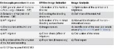
          Using an endoscopic approach, small intraoral bone chambers, which are routinely obtained during tooth extraction and implantation, provide visual in vivo access to internal bone structures. The aim of the present paper is to present a new method to quantify bone microstructure and vascularisation in vivo. Ten extraction sockets and 6 implant sites in 14 patients (6 men / 8 women) were examined by support immersion endoscopy (SIE). After tooth extraction or implant site preparation, microscopic bone analysis (MBA) was performed using short distance SIE video sequences of representative bone areas for off-line analysis with ImageJ. Quantitative assessment of the microstructure and vascularisation of the bone in dental extraction and implant sites in vivo was performed using ImageJ. MBA revealed bone morphology details such as unmineralised and mineralised areas, vascular canals and the presence of bleeding through vascular canals. Morphometric examination revealed that there was more unmineralised bone and less vascular canal area in the implant sites than in the extraction sockets.

          Related collections

          Most cited references26

          • Record: found
          • Abstract: found
          • Article: not found

          Bone classification: an objective scale of bone density using the computerized tomography scan.

          Dental implants are subject to masticatory loads of varying magnitude. Implant performance is closely related to load transmission at the bone-to-implant interface where bone quality will be highly variable. The type and architecture of bone is known to influence its load bearing capacity and it has been demonstrated that poorer quality bone is associated with higher failure rates. To date, bone classifications have only provided rough subjective methods for pre-operative assessment, which can prove unreliable. The results of an extensive analysis of computerized tomography scans using Simplant software (Columbia Scientific Inc., Columbia, MD, USA) demonstrate that an objective scale of bone density based on the Houndsfield scale, can be established and that there is a strong correlation between bone density value and subjective quality score (P = 0.002) as well as between the bone density score and the region of the mouth (P < 0.001).
            Bookmark
            • Record: found
            • Abstract: found
            • Article: not found

            Trabecular bone architecture in the pathogenesis and prevention of fracture.

            Rapid loss of trabecular bone (as after menopause) occurs by complete removal of some structural elements, leaving those that remain more widely separated and less well connected. The most likely cellular mechanism is an increase in the number of resorption cavities deep enough to lead to focal perforation of trabecular plates, either as a non-specific consequence of increased remodeling activation, or as a specific consequence (direct or indirect) of estrogen deficiency. Disruption of the connections between structural elements produces a disproportionate loss of strength, for which the increased thickness of the remaining trabeculae can only partly compensate. Consequently, the most biomechanically significant component of trabecular bone loss occurs rapidly and irreversibly. This emphasizes the importance of prevention, but no treatment except estrogen replacement is of proven efficacy in preventing estrogen-dependent bone loss. For adequate repair of structural damage after it has been allowed to occur, adding bone to existing surfaces may be insufficient, and it may be necessary to devise some means of forming new bone directly in the bone marrow cavity in order to re-establish normal connectivity.
              Bookmark
              • Record: found
              • Abstract: found
              • Article: not found

              Bone density assessments of oral implant sites using computerized tomography.

              The type and architecture of bone are considered to affect its load-bearing capacity and it has been indicated that poorer quality bone is associated with higher implant failure rates. To date, bone classifications have only provided subjective methods for pre-operative assessment, which can be considered unreliable. The aim of this study was to evaluate variations of the bone density in designated endosseous implant sites using computerized tomography. One hundred and thirty-one designated implant sites in 72 patients were utilized. Computerized tomography results indicated that bone densities may vary markedly when different areas of a designated implant site are compared. It has been observed that a difference in the bone density exists for the four regions within the oral area, with the anterior mandible yielding mean density values of 944.9+/-207 Hounsfield units (HU)>anterior maxilla, 715.8+/-190 HU>posterior mandible, 674.3+/-227 HU>posterior maxilla and 455.1+/-122 HU. Computerized tomography may be a useful tool for determining the bone density of interest areas before implant placement, and this valuable information about the bone quality provides dental practitioners to make better treatment planning regarding the implant positions.
                Bookmark

                Author and article information

                Contributors
                Role: Editor
                Journal
                PLoS One
                PLoS ONE
                plos
                plosone
                PLoS ONE
                Public Library of Science (San Francisco, CA USA )
                1932-6203
                29 December 2015
                2015
                : 10
                : 12
                : e0145767
                Affiliations
                [1 ]Department Oral and Maxillofacial Surgery, Georg-August-University Hospital, Göttingen, Germany
                [2 ]Department of Molecular Biology of Neuronal Signals, Max Planck Institute of Experimental Medicine, Göttingen, Germany
                [3 ]Research Centre in Dental Sciences (CICO), Dental School, Universidad de La Frontera, Temuco, Chile
                University of Zaragoza, SPAIN
                Author notes

                Competing Interests: The authors have declared that no competing interests exist.

                Conceived and designed the experiments: EW LM BV. Performed the experiments: EW LM BV. Analyzed the data: EW LM BV. Contributed reagents/materials/analysis tools: EW LM BV SW. Wrote the paper: EW LM BV.

                Article
                PONE-D-15-40510
                10.1371/journal.pone.0145767
                4695096
                26713617
                f2f8e089-8950-4dce-9366-6789c1accb72
                © 2015 Engelke et al

                This is an open access article distributed under the terms of the Creative Commons Attribution License, which permits unrestricted use, distribution, and reproduction in any medium, provided the original author and source are credited

                History
                : 14 September 2015
                : 8 December 2015
                Page count
                Figures: 2, Tables: 1, Pages: 9
                Funding
                The authors have no support or funding to report.
                Categories
                Research Article
                Custom metadata
                All relevant data are within the paper and its Supporting Information files.

                Uncategorized
                Uncategorized

                Comments

                Comment on this article