12
views
0
recommends
+1 Recommend
0 collections
    0
    shares
      • Record: found
      • Abstract: found
      • Article: found
      Is Open Access

      Characterization of photoreceptor degeneration in the rhodopsin P23H transgenic rat line 2 using optical coherence tomography

      research-article

      Read this article at

      Bookmark
          There is no author summary for this article yet. Authors can add summaries to their articles on ScienceOpen to make them more accessible to a non-specialist audience.

          Abstract

          Purpose

          To characterize the optical coherence tomography (OCT) appearances of photoreceptor degeneration in the rhodopsin P23H transgenic rat (line 2) in relation to the histological, ultrastructural, and electroretinography (ERG) findings.

          Materials and methods

          Homozygous rhodopsin P23H transgenic albino rats (line 2, very-slow degeneration model) were employed. Using OCT (Micron IV ®; Phoenix Research Labs, Pleasanton, CA, USA), the natural course of photoreceptor degeneration was recorded from postnatal day (P) 15 to P 287. The OCT images were qualitatively observed by comparing them to histological and ultrastructural findings at P 62 and P 169. In addition, each retinal layer was quantitatively analyzed longitudinally during degeneration, compared it to that observed in wild type Sprague-Dawley (SD) rats. The relationships between the ERG (full-field combined rod-cone response, 3.0 cds/m 2 stimulation) findings and OCT images were also analyzed.

          Results

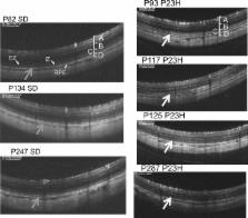
          In the qualitative study, the two layers presumably corresponding to the photoreceptor inner segment ellipsoid zone (EZ) and interdigitation zone (IZ) were identified in the P23H rat until PN day 32. However, the photoreceptor inner and outer segment (IS/OS) layer became diffusely hyperreflective on OCT after P 46, and the EZ and IZ zones could no longer be identified on OCT. In contrast, in the SD rats, the EZ and IZ were clearly distinguished until at least P 247. The ultrastructural study showed partial disarrangements of the photoreceptor outer segment discs in the P23H rats at P 62, although a light-microscopic histological study detected almost no abnormality in the outer segment. In the quantitative study, the outer retinal layer including the outer plexiform layer (OPL) and the outer nuclear layer (ONL) became significantly thinner in the P23H rats than in the SD rats after P 71. The thickness of the IS/OS layer was maintained in the P23H rats until P 130, and it became statistically thinner than in the SD rats at P 237. The longitudinal attenuation in the amplitude of the a- and b-waves of ERG was significantly correlated with the thickness of the combined OPL and ONL but not with that of the IS/OS layer.

          Conclusion

          OCT showed the degenerated photoreceptor IS/OS layer in rhodopsin P23H transgenic rats (line 2) as a diffuse hyperreflective zone, even in the early stage, with the partially disarranged and destabilized OS discs recognizable by ultrastructural assessment but not by a histological study. The amplitude of the a- and b-waves mainly depends on the thickness of the OPL and ONL layer rather than the thickness of the photoreceptor IS/OS layer in P23H rats.

          Related collections

          Most cited references27

          • Record: found
          • Abstract: found
          • Article: not found

          Anatomical correlates to the bands seen in the outer retina by optical coherence tomography: literature review and model.

          To evaluate the validity of commonly used anatomical designations for the four hyperreflective outer retinal bands seen in current-generation optical coherence tomography, a scale model of outer retinal morphology was created using published information for direct comparison with optical coherence tomography scans. Articles and books concerning histology of the outer retina from 1900 until 2009 were evaluated, and data were used to create a scale model drawing. Boundaries between outer retinal tissue compartments described by the model were compared with intensity variations of representative spectral-domain optical coherence tomography scans using longitudinal reflectance profiles to determine the region of origin of the hyperreflective outer retinal bands. This analysis showed a high likelihood that the spectral-domain optical coherence tomography bands attributed to the external limiting membrane (the first, innermost band) and to the retinal pigment epithelium (the fourth, outermost band) are correctly attributed. Comparative analysis showed that the second band, often attributed to the boundary between inner and outer segments of the photoreceptors, actually aligns with the ellipsoid portion of the inner segments. The third band corresponded to an ensheathment of the cone outer segments by apical processes of the retinal pigment epithelium in a structure known as the contact cylinder. Anatomical attributions and subsequent pathophysiologic assessments pertaining to the second and third outer retinal hyperreflective bands may not be correct. This analysis has identified testable hypotheses for the actual correlates of the second and third bands. Nonretinal pigment epithelium contributions to the fourth band (e.g., Bruch membrane) remain to be determined.
            Bookmark
            • Record: found
            • Abstract: found
            • Article: not found

            INHERITED RETINAL DYSTROPHY IN THE RAT

            Retinal dystrophies, known in man, dog, mouse, and rat, involve progressive loss of photoreceptor cells with onset during or soon after the developmental period. Functional (electroretinogram), chemical (rhodopsin analyses) and morphological (light and electron microscopy) data obtained in the rat indicated two main processes: (a) overproduction of rhodopsin and an associated abnormal lamellar tissue component, (b) progressive loss of photoreceptor cells. The first abnormality recognized was the appearance of swirling sheets or bundles of extracellular lamellae between normally developing retinal rods and pigment epithelium; membrane thickness and spacing resembled that in normal outer segments. Rhodopsin content reached twice normal values, was present in both rods and extracellular lamellae, and was qualitatively normal, judged by absorption maximum and products of bleaching. Photoreceptors attained virtually adult form and ERG function. Then rod inner segments and nuclei began degenerating; the ERG lost sensitivity and showed selective depression of the a-wave at high luminances. Outer segments and lamellae gradually degenerated and rhodopsin content decreased. No phagocytosis was seen, though pigment cells partially dedifferentiated and many migrated through the outer segment-debris zone toward the retina. Eventually photoreceptor cells and the b-wave of the ERG entirely disappeared. Rats kept in darkness retained electrical activity, rhodopsin content, rod structure, and extracellular lamellae longer than litter mates in light.
              Bookmark
              • Record: found
              • Abstract: found
              • Article: not found

              A rhodopsin mutant linked to autosomal dominant retinitis pigmentosa is prone to aggregate and interacts with the ubiquitin proteasome system.

              The inherited retinal degenerations are typified by retinitis pigmentosa (RP), a heterogeneous group of inherited disorders that causes the destruction of photoreceptor cells, the retinal pigmented epithelium, and choroid. This group of blinding conditions affects over 1.5 million persons worldwide. Approximately 30-40% of human autosomal dominant (AD) RP is caused by dominantly inherited missense mutations in the rhodopsin gene. Here we show that P23H, the most frequent RP mutation in American patients, renders rhodopsin extremely prone to form high molecular weight oligomeric species in the cytoplasm of transfected cells. Aggregated P23H accumulates in aggresomes, which are pericentriolar inclusion bodies that require an intact microtubule cytoskeleton to form. Using fluorescence resonance energy transfer (FRET), we observe that P23H aggregates in the cytoplasm even at extremely low expression levels. Our data show that the P23H mutation destabilizes the protein and targets it for degradation by the ubiquitin proteasome system. P23H is stabilized by proteasome inhibitors and by co-expression of a dominant negative form of ubiquitin. We show that expression of P23H, but not wild-type rhodopsin, results in a generalized impairment of the ubiquitin proteasome system, suggesting a mechanism for photoreceptor degeneration that links RP to a broad class of neurodegenerative diseases.
                Bookmark

                Author and article information

                Contributors
                Role: Data curationRole: InvestigationRole: MethodologyRole: Writing – original draft
                Role: Data curationRole: MethodologyRole: Writing – review & editing
                Role: Data curationRole: InvestigationRole: Writing – review & editing
                Role: Data curationRole: InvestigationRole: Writing – review & editing
                Role: Data curationRole: Formal analysisRole: SupervisionRole: VisualizationRole: Writing – review & editing
                Role: Formal analysisRole: Funding acquisitionRole: Project administrationRole: SupervisionRole: Writing – review & editing
                Role: Editor
                Journal
                PLoS One
                PLoS ONE
                plos
                plosone
                PLoS ONE
                Public Library of Science (San Francisco, CA USA )
                1932-6203
                9 March 2018
                2018
                : 13
                : 3
                : e0193778
                Affiliations
                [1 ] Department of Ophthalmology, Hirosaki University Graduate School of Medicine, Hirosaki, Japan
                [2 ] Department of Ophthalmology, Tohoku University Graduate School of Medicine, Sendai, Japan
                University of Florida, UNITED STATES
                Author notes

                Competing Interests: Research fundings were from Pfizer, Santen, Alcon, Novartis, K-vision, AMO. This does not alter our adherence to PLOS ONE policies on sharing data and materials.

                Author information
                http://orcid.org/0000-0001-6095-5712
                Article
                PONE-D-17-39967
                10.1371/journal.pone.0193778
                5844545
                29522537
                f316f25b-3092-468f-ae93-666ade5f4fc1
                © 2018 Monai et al

                This is an open access article distributed under the terms of the Creative Commons Attribution License, which permits unrestricted use, distribution, and reproduction in any medium, provided the original author and source are credited.

                History
                : 22 November 2017
                : 16 February 2018
                Page count
                Figures: 9, Tables: 2, Pages: 17
                Funding
                Funded by: funder-id http://dx.doi.org/10.13039/501100000646, Japan Society for the Promotion of Science London;
                Award ID: C-16K11313
                Award Recipient :
                Funded by: funder-id http://dx.doi.org/10.13039/501100000646, Japan Society for the Promotion of Science London;
                Award ID: B-17K16954
                Award Recipient :
                This study was supported in part by a Grant-in Aid for Scientific Research, C-16K11313 (MN) from Japan Society for the Promotion of Science, http://www.jsps.go.jp/, and a Grant-in Aid for Young Scientists, B-17K16954 (NM) from Japan Society for the Promotion of Science, http://www.jsps.go.jp/. Research Fundings from Pfizer, Santen, Alcon, Novartis, K-vision, AMO also supported this study. The funders had no role in study design, data collection and analysis, decision to publish, or preparation of the manuscript.
                Categories
                Research Article
                Biology and Life Sciences
                Cell Biology
                Cellular Types
                Animal Cells
                Neurons
                Afferent Neurons
                Photoreceptors
                Biology and Life Sciences
                Neuroscience
                Cellular Neuroscience
                Neurons
                Afferent Neurons
                Photoreceptors
                Biology and Life Sciences
                Neuroscience
                Sensory Perception
                Sensory Receptors
                Photoreceptors
                Biology and Life Sciences
                Psychology
                Sensory Perception
                Sensory Receptors
                Photoreceptors
                Social Sciences
                Psychology
                Sensory Perception
                Sensory Receptors
                Photoreceptors
                Biology and Life Sciences
                Cell Biology
                Signal Transduction
                Sensory Receptors
                Photoreceptors
                Medicine and Health Sciences
                Diagnostic Medicine
                Diagnostic Radiology
                Tomography
                Research and Analysis Methods
                Imaging Techniques
                Diagnostic Radiology
                Tomography
                Medicine and Health Sciences
                Radiology and Imaging
                Diagnostic Radiology
                Tomography
                Biology and Life Sciences
                Anatomy
                Histology
                Medicine and Health Sciences
                Anatomy
                Histology
                Medicine and Health Sciences
                Ophthalmology
                Retinal Disorders
                Retinal Degeneration
                Biology and Life Sciences
                Anatomy
                Ocular System
                Ocular Anatomy
                Retina
                Medicine and Health Sciences
                Anatomy
                Ocular System
                Ocular Anatomy
                Retina
                Biology and Life Sciences
                Anatomy
                Head
                Eyes
                Medicine and Health Sciences
                Anatomy
                Head
                Eyes
                Biology and Life Sciences
                Anatomy
                Ocular System
                Eyes
                Medicine and Health Sciences
                Anatomy
                Ocular System
                Eyes
                Medicine and Health Sciences
                Ophthalmology
                Retinal Disorders
                Retinitis
                Retinitis Pigmentosa
                Biology and Life Sciences
                Genetics
                Mutation
                Point Mutation
                Custom metadata
                All relevant data are within the paper and its Supporting Information files.

                Uncategorized
                Uncategorized

                Comments

                Comment on this article