35
views
0
recommends
+1 Recommend
0 collections
    0
    shares
      • Record: found
      • Abstract: found
      • Article: found
      Is Open Access

      Mineral-Doped Poly(L-lactide) Acid Scaffolds Enriched with Exosomes Improve Osteogenic Commitment of Human Adipose-Derived Mesenchymal Stem Cells

      research-article

      Read this article at

      Bookmark
          There is no author summary for this article yet. Authors can add summaries to their articles on ScienceOpen to make them more accessible to a non-specialist audience.

          Abstract

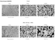
          Exosomes derived from mesenchymal stem cells are extracellular vesicles released to facilitate cell communication and function. Recently, polylactic acid (PLA), calcium silicates (CaSi), and dicalcium phosphate dihydrate (DCPD) have been used to produce bioresorbable functional mineral-doped porous scaffolds-through thermally induced phase separation technique, as materials for bone regeneration. The aim of this study was to investigate the effect of mineral-doped PLA-based porous scaffolds enriched with exosome vesicles (EVs) on osteogenic commitment of human adipose mesenchymal stem cells (hAD-MSCs). Two different mineral-doped scaffolds were produced: PLA-10CaSi-10DCPD and PLA-5CaSi-5DCPD. Scaffolds surface micromorphology was investigated by ESEM-EDX before and after 28 days immersion in simulated body fluid (HBSS). Exosomes were deposited on the surface of the scaffolds and the effect of exosome-enriched scaffolds on osteogenic commitment of hAD-MSCs cultured in proximity of the scaffolds has been evaluated by real time PCR. In addition, the biocompatibility was evaluated by direct-contact seeding hAD-MSCs on scaffolds surface-using MTT viability test. In both formulations, ESEM showed pores similar in shape (circular and elliptic) and size (from 10–30 µm diameter). The porosity of the scaffolds decreased after 28 days immersion in simulated body fluid. Mineral-doped scaffolds showed a dynamic surface and created a suitable bone-forming microenvironment. The presence of the mineral fillers increased the osteogenic commitment of hAD-MSCs. Exosomes were easily entrapped on the surface of the scaffolds and their presence improved gene expression of major markers of osteogenesis such as collagen type I, osteopontin, osteonectin, osteocalcin. The experimental scaffolds enriched with exosomes, in particular PLA-10CaSi-10DCPD, increased the osteogenic commitment of MSCs. In conclusion, the enrichment of bioresorbable functional scaffolds with exosomes is confirmed as a potential strategy to improve bone regeneration procedures.

          Related collections

          Most cited references71

          • Record: found
          • Abstract: found
          • Article: not found

          The biology of fracture healing.

          The biology of fracture healing is a complex biological process that follows specific regenerative patterns and involves changes in the expression of several thousand genes. Although there is still much to be learned to fully comprehend the pathways of bone regeneration, the over-all pathways of both the anatomical and biochemical events have been thoroughly investigated. These efforts have provided a general understanding of how fracture healing occurs. Following the initial trauma, bone heals by either direct intramembranous or indirect fracture healing, which consists of both intramembranous and endochondral bone formation. The most common pathway is indirect healing, since direct bone healing requires an anatomical reduction and rigidly stable conditions, commonly only obtained by open reduction and internal fixation. However, when such conditions are achieved, the direct healing cascade allows the bone structure to immediately regenerate anatomical lamellar bone and the Haversian systems without any remodelling steps necessary. In all other non-stable conditions, bone healing follows a specific biological pathway. It involves an acute inflammatory response including the production and release of several important molecules, and the recruitment of mesenchymal stem cells in order to generate a primary cartilaginous callus. This primary callus later undergoes revascularisation and calcification, and is finally remodelled to fully restore a normal bone structure. In this article we summarise the basic biology of fracture healing. Copyright © 2011 Elsevier Ltd. All rights reserved.
            Bookmark
            • Record: found
            • Abstract: found
            • Article: not found

            Clinical applications of mineral trioxide aggregate.

            An experimental material, mineral trioxide aggregate (MTA), has recently been investigated as a potential alternative restorative material to the presently used materials in endodontics. Several in vitro and in vivo studies have shown that MTA prevents microleakage, is biocompatible, and promotes regeneration of the original tissues when it is placed in contact with the dental pulp or periradicular tissues. This article describes the clinical procedures for application of MTA in capping of pulps with reversible pulpitis, apexification, repair of root perforations nonsurgically and surgically, as well as its use as a root-end filling material.
              Bookmark
              • Record: found
              • Abstract: found
              • Article: not found

              MicroRNA control of bone formation and homeostasis.

              MicroRNAs (miRNAs) repress cellular protein levels to provide a sophisticated parameter of gene regulation that coordinates a broad spectrum of biological processes. Bone organogenesis is a complex process involving the differentiation and crosstalk of multiple cell types for formation and remodeling of the skeleton. Inhibition of mRNA translation by miRNAs has emerged as an important regulator of developmental osteogenic signaling pathways, osteoblast growth and differentiation, osteoclast-mediated bone resorption activity and bone homeostasis in the adult skeleton. miRNAs control multiple layers of gene regulation for bone development and postnatal functions, from the initial response of stem/progenitor cells to the structural and metabolic activity of the mature tissue. This Review brings into focus an emerging concept of bone-regulating miRNAs, the evidence for which has been gathered largely from in vivo mouse models and in vitro studies in human and mouse skeletal cell populations. Characterization of miRNAs that operate through tissue-specific transcription factors in osteoblast and osteoclast lineage cells, as well as intricate feedforward and reverse loops, has provided novel insights into the supervision of signaling pathways and regulatory networks controlling normal bone formation and turnover. The current knowledge of miRNAs characteristic of human pathologic disorders of the skeleton is presented with a future goal towards translational studies.
                Bookmark

                Author and article information

                Journal
                Nanomaterials (Basel)
                Nanomaterials (Basel)
                nanomaterials
                Nanomaterials
                MDPI
                2079-4991
                29 February 2020
                March 2020
                : 10
                : 3
                : 432
                Affiliations
                [1 ]Laboratory of Biomaterials and Oral Pathology, School of Dentistry, Department of Biomedical and Neuromotor Sciences, University of Bologna, 40125 Bologna, Italy; fausto.zamparini2@ 123456unibo.it (F.Z.);
                [2 ]Medical Sciences Department, University of Ferrara, 44100 Ferrara, Italy
                [3 ]Department of Civil, Chemical, Environmental and Materials Engineering, University of Bologna, 40136 Bologna, Italy
                [4 ]Department of Metallurgical and Materials Engineering, Middle East Technical University (METU), 06800 Ankara, Turkey
                [5 ]Biomedical Engineering Program, METU, 06800 Ankara, Turkey
                [6 ]BIOMATEN, Center of Excellence in Biomaterials and Tissue Engineering, METU, 06800 Ankara, Turkey
                [7 ]Cellular Signaling Laboratory, Institute of Human Anatomy, Department of Biomedical and Neuromotor Sciences, University of Bologna, 40126 Bologna, Italy
                [8 ]Endodontic Clinical Section, School of Dentistry, Department of Biomedical and Neuromotor Sciences, University of Bologna, 40125 Bologna, Italy
                Author notes
                Author information
                https://orcid.org/0000-0001-7793-6227
                https://orcid.org/0000-0002-0071-4463
                https://orcid.org/0000-0002-4513-8527
                https://orcid.org/0000-0002-7764-4539
                https://orcid.org/0000-0002-4779-4456
                Article
                nanomaterials-10-00432
                10.3390/nano10030432
                7153699
                32121340
                f63ebcac-2975-4619-a3d4-13e703601b5e
                © 2020 by the authors.

                Licensee MDPI, Basel, Switzerland. This article is an open access article distributed under the terms and conditions of the Creative Commons Attribution (CC BY) license ( http://creativecommons.org/licenses/by/4.0/).

                History
                : 05 December 2019
                : 22 February 2020
                Categories
                Article

                exosomes,human adipose mesenchymal stem cells (had-mscs), polylactic acid scaffold,calcium silicates,exosome enriched scaffolds,regenerative bone healing,green bioplastics,bioresorbable scaffold,natural nanovesicles,nanodelivery,personalized regenerative medicine

                Comments

                Comment on this article