35
views
0
recommends
+1 Recommend
0 collections
    0
    shares
      • Record: found
      • Abstract: found
      • Article: found
      Is Open Access

      Human Gingival Fibroblasts Display a Non-Fibrotic Phenotype Distinct from Skin Fibroblasts in Three-Dimensional Cultures

      research-article

      Read this article at

      Bookmark
          There is no author summary for this article yet. Authors can add summaries to their articles on ScienceOpen to make them more accessible to a non-specialist audience.

          Abstract

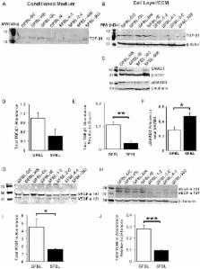
          Scar formation following skin injury can be a major psychosocial and physiological problem. However, the mechanisms of scar formation are still not completely understood. Previous studies have shown that wound healing in oral mucosa is faster, associates with a reduced inflammatory response and results to significantly reduced scar formation compared with skin wounds. In the present study, we hypothesized that oral mucosal fibroblasts from human gingiva are inherently distinct from fibroblasts from breast and abdominal skin, two areas prone to excessive scar formation, which may contribute to the preferential wound healing outcome in gingiva. To this end, we compared the phenotype of human gingival and skin fibroblasts cultured in in vivo-like three-dimensional (3D) cultures that mimic the cells' natural extracellular matrix (ECM) niche. To establish 3D cultures, five parallel fibroblast lines from human gingiva (GFBLs) and breast skin (SFBLs) were seeded in high density, and cultured for up to 21 days in serum and ascorbic acid containing medium to induce expression of wound-healing transcriptome and ECM deposition. Cell proliferation, morphology, phenotype and expression of wound healing and scar related genes were analyzed by real-time RT-PCR, Western blotting and immunocytochemical methods. The expression of a set of genes was also studied in three parallel lines of human abdominal SFBLs. Findings showed that GFBLs displayed morphologically distinct organization of the 3D cultures and proliferated faster than SFBLs. GFBLs expressed elevated levels of molecules involved in regulation of inflammation and ECM remodeling (MMPs) while SFBLs showed significantly higher expression of TGF-β signaling, ECM and myofibroblast and cell contractility-related genes. Thus, GFBLs display an inherent phenotype conducive for fast resolution of inflammation and ECM remodeling, characteristic for scar-free wound healing, while SFBLs have a profibrotic, scar-prone phenotype.

          Related collections

          Most cited references57

          • Record: found
          • Abstract: found
          • Article: not found

          The transcriptional program in the response of human fibroblasts to serum.

          The temporal program of gene expression during a model physiological response of human cells, the response of fibroblasts to serum, was explored with a complementary DNA microarray representing about 8600 different human genes. Genes could be clustered into groups on the basis of their temporal patterns of expression in this program. Many features of the transcriptional program appeared to be related to the physiology of wound repair, suggesting that fibroblasts play a larger and richer role in this complex multicellular response than had previously been appreciated.
            Bookmark
            • Record: found
            • Abstract: not found
            • Article: not found

            SPARC, a matricellular protein that functions in cellular differentiation and tissue response to injury.

              Bookmark
              • Record: found
              • Abstract: found
              • Article: not found

              Healing and hurting: molecular mechanisms, functions, and pathologies of cellular senescence.

              Cellular senescence is a proliferation arrest that is typically irreversible and caused by various cellular stresses, including excess rounds of cell division and cancer-causing genetic alterations. Senescence actively contributes to a tissue-level response to tissue wounding and incipient cancer, healing the tissue and suppressing tumor formation. However, in the long term, the same senescence program may hurt the tissue, thereby contributing to tissue aging. Tumor suppression, wound healing, and aging are each associated with inflammation, and here it is proposed that cellular senescence contributes to a "nonimmune cell" component of the tissue inflammatory response.
                Bookmark

                Author and article information

                Contributors
                Role: Editor
                Journal
                PLoS One
                PLoS ONE
                plos
                plosone
                PLoS ONE
                Public Library of Science (San Francisco, USA )
                1932-6203
                2014
                7 March 2014
                : 9
                : 3
                : e90715
                Affiliations
                [1]Department of Oral Biological and Medical Sciences, Faculty of Dentistry, University of British Columbia, Vancouver, Canada
                University of Bergen, Norway
                Author notes

                Competing Interests: The authors have declared that no competing interests exist.

                Conceived and designed the experiments: WM GJ DO GC BK HL LH. Performed the experiments: WM GJ DO GC BK. Analyzed the data: WM GJ DO GC BK HL LH. Contributed reagents/materials/analysis tools: HL LH. Wrote the paper: WM GJ DO GC BK HL LH.

                Article
                PONE-D-13-46665
                10.1371/journal.pone.0090715
                3946595
                24608113
                f7111cb9-85e3-418a-af25-96cac071d8e3
                Copyright @ 2014

                This is an open-access article distributed under the terms of the Creative Commons Attribution License, which permits unrestricted use, distribution, and reproduction in any medium, provided the original author and source are credited.

                History
                : 6 November 2013
                : 5 February 2014
                Page count
                Pages: 20
                Funding
                The study was supported by Canadian Institutes of Health Research (CIHR, MOP 77550), University of British Columbia CIHR Skin Research Training Centre (SRTC) Award, and by The Faculty of Dentistry, University of British Columbia, Undergraduate Student Research Awards. The funders had no role in study design, data collection and analysis, decision to publish, or preparation of the manuscript.
                Categories
                Research Article
                Biology
                Biochemistry
                Enzymes
                Enzyme Classes
                Collagenase
                Molecular cell biology
                Cellular Types
                Connective Tissue Cells
                Extracellular Matrix
                Connective Tissue
                Extracellular Matrix Composition
                Integrins
                Signal transduction
                Signaling cascades
                TGF-beta signaling cascade
                Gene Expression
                Medicine
                Anatomy and Physiology
                Skin
                Dermatology
                Oral Medicine
                Oral Diseases

                Uncategorized
                Uncategorized

                Comments

                Comment on this article