25
views
0
recommends
+1 Recommend
0 collections
    0
    shares
      • Record: found
      • Abstract: found
      • Article: found
      Is Open Access

      Whole-brain background-suppressed pCASL MRI with 1D-accelerated 3D RARE Stack-Of-Spirals readout

      research-article

      Read this article at

      Bookmark
          There is no author summary for this article yet. Authors can add summaries to their articles on ScienceOpen to make them more accessible to a non-specialist audience.

          Abstract

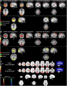
          Arterial Spin Labeled (ASL) perfusion MRI enables non-invasive, quantitative measurements of tissue perfusion, and has a broad range of applications including brain functional imaging. However, ASL suffers from low signal-to-noise ratio (SNR), limiting image resolution. Acquisitions using 3D readouts are optimal for background-suppression of static signals, but can be SAR intensive and typically suffer from through-plane blurring. In this study, we investigated the use of accelerated 3D readouts to obtain whole-brain, high-SNR ASL perfusion maps and reduce SAR deposition. Parallel imaging was implemented along the partition-encoding direction in a pseudo-continuous ASL sequence with background-suppression and 3D RARE Stack-Of-Spirals readout, and its performance was evaluated in three small cohorts. First, both non-accelerated and two-fold accelerated single-shot versions of the sequence were evaluated in healthy volunteers during a motor-photic task, and the performance was compared in terms of temporal SNR, GM-WM contrast, and statistical significance of the detected activation. Secondly, single-shot 1D-accelerated imaging was compared to a two-shot accelerated version to assess benefits of SNR and spatial resolution for applications in which temporal resolution is not paramount. Third, the efficacy of this approach in clinical populations was assessed by applying the single-shot 1D-accelerated version to a larger cohort of elderly volunteers. Accelerated data demonstrated the ability to detect functional activation at the subject level, including cerebellar activity, without loss in the perfusion signal temporal stability and the statistical power of the activations. The use of acceleration also resulted in increased GM-WM contrast, likely due to reduced through-plane partial volume effects, that were further attenuated with the use of two-shot readouts. In a clinical cohort, image quality remained excellent, and expected effects of age and sex on cerebral blood flow could be detected. The sequence is freely available upon request for academic use and could benefit a broad range of cognitive and clinical neuroscience research.

          Related collections

          Most cited references19

          • Record: found
          • Abstract: found
          • Article: not found

          Magnetic resonance imaging of perfusion using spin inversion of arterial water.

          A technique has been developed for proton magnetic resonance imaging (MRI) of perfusion, using water as a freely diffusable tracer, and its application to the measurement of cerebral blood flow (CBF) in the rat is demonstrated. The method involves labeling the inflowing water proton spins in the arterial blood by inverting them continuously at the neck region and observing the effects of inversion on the intensity of brain MRI. Solution to the Bloch equations, modified to include the effects of flow, allows regional perfusion rates to be measured from an image with spin inversion, a control image, and a T1 image. Continuous spin inversion labeling the arterial blood water was accomplished, using principles of adiabatic fast passage by applying continuous-wave radiofrequency power in the presence of a magnetic field gradient in the direction of arterial flow. In the detection slice used to measure perfusion, whole brain CBF averaged 1.39 +/- 0.19 ml.g-1.min-1 (mean +/- SEM, n = 5). The technique's sensitivity to changes in CBF was measured by using graded hypercarbia, a condition that is known to increase brain perfusion. CBF vs. pCO2 data yield a best-fit straight line described by CBF (ml.g-1.min-1) = 0.052pCO2 (mm Hg) - 0.173, in excellent agreement with values in the literature. Finally, perfusion images of a freeze-injured rat brain have been obtained, demonstrating the technique's ability to detect regional abnormalities in perfusion.
            Bookmark
            • Record: found
            • Abstract: not found
            • Article: not found

            Some windows with very good sidelobe behavior

            A. Nuttall (1981)
              Bookmark
              • Record: found
              • Abstract: found
              • Article: not found

              Behind the scenes of functional brain imaging: a historical and physiological perspective.

              M Raichle (1998)
              At the forefront of cognitive neuroscience research in normal humans are the new techniques of functional brain imaging: positron emission tomography and magnetic resonance imaging. The signal used by positron emission tomography is based on the fact that changes in the cellular activity of the brain of normal, awake humans and laboratory animals are accompanied almost invariably by changes in local blood flow. This robust, empirical relationship has fascinated scientists for well over a hundred years. Because the changes in blood flow are accompanied by lesser changes in oxygen consumption, local changes in brain oxygen content occur at the sites of activation and provide the basis for the signal used by magnetic resonance imaging. The biological basis for these signals is now an area of intense research stimulated by the interest in these tools for cognitive neuroscience research.
                Bookmark

                Author and article information

                Contributors
                Role: ConceptualizationRole: Formal analysisRole: InvestigationRole: SoftwareRole: Writing – original draftRole: Writing – review & editing
                Role: ConceptualizationRole: Funding acquisitionRole: Writing – review & editing
                Role: Writing – review & editing
                Role: Funding acquisitionRole: Writing – review & editing
                Role: ConceptualizationRole: Writing – review & editing
                Role: ConceptualizationRole: Funding acquisitionRole: SupervisionRole: Writing – review & editing
                Role: Editor
                Journal
                PLoS One
                PLoS ONE
                plos
                plosone
                PLoS ONE
                Public Library of Science (San Francisco, CA USA )
                1932-6203
                24 August 2017
                2017
                : 12
                : 8
                : e0183762
                Affiliations
                [1 ] Department of Neurology, University of Pennsylvania, Philadelphia, Pennsylvania, United States of America
                [2 ] Department of Radiology, University of Pennsylvania, Philadelphia, Pennsylvania, United States of America
                [3 ] Department of Radiology, Temple University, Philadelphia, Pennsylvania, United States of America
                [4 ] Center for Cognition and Brain Disorder, Hangzhou Normal University, Hangzhou, Zhejiang Province, China
                [5 ] Department of Radiology, Clínica Universidad de Navarra, Pamplona, Navarra, Spain
                Lee Kong Chian School of Medicine, SINGAPORE
                Author notes

                Competing Interests: The authors have declared that no competing interests exist.

                Article
                PONE-D-17-03909
                10.1371/journal.pone.0183762
                5570334
                28837640
                f926ee12-c007-4ea0-9594-3c51b51a9f72
                © 2017 Vidorreta et al

                This is an open access article distributed under the terms of the Creative Commons Attribution License, which permits unrestricted use, distribution, and reproduction in any medium, provided the original author and source are credited.

                History
                : 30 January 2017
                : 10 August 2017
                Page count
                Figures: 6, Tables: 2, Pages: 16
                Funding
                Funded by: funder-id http://dx.doi.org/10.13039/100000009, Foundation for the National Institutes of Health;
                Award ID: P41EB015893
                Award Recipient :
                Funded by: funder-id http://dx.doi.org/10.13039/100000009, Foundation for the National Institutes of Health;
                Award ID: MH080729
                Award Recipient :
                Funded by: funder-id http://dx.doi.org/10.13039/501100001809, National Natural Science Foundation of China;
                Award ID: 81471644
                Award Recipient :
                Funded by: funder-id http://dx.doi.org/10.13039/100000009, Foundation for the National Institutes of Health;
                Award ID: R01 AG040271
                Award Recipient :
                This work has was supported by the National Institutes of Health ( http://www.nih.gov/), grants no. P41EB015893, MH080729, and R01 AG040271, by National Natural Science Foundation of China ( http://www.nsfc.gov.cn/), grant no. 81471644, and Hangzhou Innovation Seed Fund. The funders had no role in study design, data collection and analysis, decision to publish, or preparation of the manuscript.
                Categories
                Research Article
                Biology and Life Sciences
                Neuroscience
                Brain Mapping
                Functional Magnetic Resonance Imaging
                Arterial Spin Labelling
                Medicine and Health Sciences
                Diagnostic Medicine
                Diagnostic Radiology
                Magnetic Resonance Imaging
                Functional Magnetic Resonance Imaging
                Arterial Spin Labelling
                Research and Analysis Methods
                Imaging Techniques
                Diagnostic Radiology
                Magnetic Resonance Imaging
                Functional Magnetic Resonance Imaging
                Arterial Spin Labelling
                Medicine and Health Sciences
                Radiology and Imaging
                Diagnostic Radiology
                Magnetic Resonance Imaging
                Functional Magnetic Resonance Imaging
                Arterial Spin Labelling
                Research and Analysis Methods
                Imaging Techniques
                Neuroimaging
                Functional Magnetic Resonance Imaging
                Arterial Spin Labelling
                Biology and Life Sciences
                Neuroscience
                Neuroimaging
                Functional Magnetic Resonance Imaging
                Arterial Spin Labelling
                Medicine and Health Sciences
                Diagnostic Medicine
                Diagnostic Radiology
                Magnetic Resonance Imaging
                Research and Analysis Methods
                Imaging Techniques
                Diagnostic Radiology
                Magnetic Resonance Imaging
                Medicine and Health Sciences
                Radiology and Imaging
                Diagnostic Radiology
                Magnetic Resonance Imaging
                Medicine and Health Sciences
                Geriatrics
                People and Places
                Population Groupings
                Age Groups
                Elderly
                Research and Analysis Methods
                Bioassays and Physiological Analysis
                Cardiovascular Analysis
                Cerebral Blood Flow Assay
                Computer and Information Sciences
                Data Acquisition
                Computer and Information Sciences
                Information Theory
                Background Signal Noise
                Engineering and Technology
                Signal Processing
                Background Signal Noise
                Research and Analysis Methods
                Imaging Techniques
                Custom metadata
                All data are available from the OpenfMRI database ( https://openfmri.org), accession numbers ds000234, ds000235 and ds000236.

                Uncategorized
                Uncategorized

                Comments

                Comment on this article