21
views
0
recommends
+1 Recommend
0 collections
    0
    shares
      • Record: found
      • Abstract: found
      • Article: not found

      Breed Differences in Natriuretic Peptides in Healthy Dogs

      research-article

      Read this article at

      ScienceOpenPublisherPMC
      Bookmark
          There is no author summary for this article yet. Authors can add summaries to their articles on ScienceOpen to make them more accessible to a non-specialist audience.

          Abstract

          Background

          Measurement of plasma concentration of natriuretic peptides ( NPs) is suggested to be of value in diagnosis of cardiac disease in dogs, but many factors other than cardiac status may influence their concentrations. Dog breed potentially is 1 such factor.

          Objective

          To investigate breed variation in plasma concentrations of pro‐atrial natriuretic peptide 31‐67 (pro ANP 31‐67) and N‐terminal B‐type natriuretic peptide ( NT‐pro BNP) in healthy dogs.

          Animals

          535 healthy, privately owned dogs of 9 breeds were examined at 5 centers as part of the European Union ( EU) LUPA project.

          Methods

          Absence of cardiovascular disease or other clinically relevant organ‐related or systemic disease was ensured by thorough clinical investigation. Plasma concentrations of pro ANP 31‐67 and NT‐pro BNP were measured by commercially available ELISA assays.

          Results

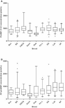
          Overall significant breed differences were found in pro ANP 31‐67 ( P < .0001) and NT‐pro BNP ( P < .0001) concentrations. Pair‐wise comparisons between breeds differed in approximately 50% of comparisons for pro ANP 31‐67 as well as NT‐pro BNP concentrations, both when including all centers and within each center. Interquartile range was large for many breeds, especially for NT‐pro BNP. Among included breeds, Labrador Retrievers and Newfoundlands had highest median NT‐pro BNP concentrations with concentrations 3 times as high as those of Dachshunds. German Shepherds and Cavalier King Charles Spaniels had the highest median pro ANP 31‐67 concentrations, twice the median concentration in Doberman Pinschers.

          Conclusions and Clinical Importance

          Considerable interbreed variation in plasma NP concentrations was found in healthy dogs. Intrabreed variation was large in several breeds, especially for NT‐pro BNP. Additional studies are needed to establish breed‐specific reference ranges.

          Related collections

          Most cited references48

          • Record: found
          • Abstract: found
          • Article: not found

          State of the art: using natriuretic peptide levels in clinical practice.

          Natriuretic peptide (NP) levels (B-type natriuretic peptide (BNP) and N-terminal proBNP) are now widely used in clinical practice and cardiovascular research throughout the world and have been incorporated into most national and international cardiovascular guidelines for heart failure. The role of NP levels in state-of-the-art clinical practice is evolving rapidly. This paper reviews and highlights ten key messages to clinicians: 1) NP levels are quantitative plasma biomarkers of heart failure (HF). 2) NP levels are accurate in the diagnosis of HF. 3) NP levels may help risk stratify emergency department (ED) patients with regard to the need for hospital admission or direct ED discharge. 4) NP levels help improve patient management and reduce total treatment costs in patients with acute dyspnoea. 5) NP levels at the time of admission are powerful predictors of outcome in predicting death and re-hospitalisation in HF patients. 6) NP levels at discharge aid in risk stratification of the HF patient. 7) NP-guided therapy may improve morbidity and/or mortality in chronic HF. 8) The combination of NP levels together with symptoms, signs and weight gain assists in the assessment of clinical decompensation in HF. 9) NP levels can accelerate accurate diagnosis of heart failure presenting in primary care. 10) NP levels may be helpful to screen for asymptomatic left ventricular dysfunction in high-risk patients.
            Bookmark
            • Record: found
            • Abstract: found
            • Article: not found

            Recommendations for standards in transthoracic two-dimensional echocardiography in the dog and cat. Echocardiography Committee of the Specialty of Cardiology, American College of Veterinary Internal Medicine.

            Recommendations are presented for standardized imaging planes and display conventions for two-dimensional echocardiography in the dog and cat. Three transducer locations ("windows") provide access to consistent imaging planes: the right parasternal location, the left caudal (apical) parasternal location, and the left cranial parasternal location. Recommendations for image display orientations are very similar to those for comparable human cardiac images, with the heart base or cranial aspect of the heart displayed to the examiner's right on the video display. From the right parasternal location, standard views include a long-axis four-chamber view and a long-axis left ventricular outflow view, and short-axis views at the levels of the left ventricular apex, papillary muscles, chordae tendineae, mitral valve, aortic valve, and pulmonary arteries. From the left caudal (apical) location, standard views include long-axis two-chamber and four-chamber views. From the left cranial parasternal location, standard views include a long-axis view of the left ventricular outflow tract and ascending aorta (with variations to image the right atrium and tricuspid valve, and the pulmonary valve and pulmonary artery), and a short-axis view of the aortic root encircled by the right heart. These images are presented by means of idealized line drawings. Adoption of these standards should facilitate consistent performance, recording, teaching, and communicating results of studies obtained by two-dimensional echocardiography.
              Bookmark
              • Record: found
              • Abstract: found
              • Article: not found

              Localization and mechanism of secretion of B-type natriuretic peptide in comparison with those of A-type natriuretic peptide in normal subjects and patients with heart failure.

              B-type or brain natriuretic peptide (BNP) is a novel natriuretic peptide secreted from the heart that forms a peptide family with A-type or atrial natriuretic peptide (ANP), and its plasma level has been shown to be increased in patients with congestive heart failure. This study was designed to examine the sources and mechanisms of the secretion of BNP in comparison with those of ANP in control subjects and in patients with heart failure. We measured the plasma levels of BNP as well as ANP in 16 patients with dilated cardiomyopathy (11 men and 5 women; mean age, 59 years) and 18 control subjects (9 men and 9 women; mean age, 54 years) by sampling blood from the femoral vein, the aortic root, the anterior interventricular vein (AIV), and the coronary sinus using the newly developed immunoradiometric assay systems. In the control subjects, there was no significant difference in the plasma ANP level between the aortic root and the AIV (24.0 +/- 5.2 pg/mL versus 32.2 +/- 17.0 pg/mL), but there was a highly significant step-up of the level between the AIV and the coronary sinus (32.2 +/- 17.0 pg/mL versus 371.4 +/- 111.1 pg/mL, P < .001). In contrast, there was a significant step-up of the plasma BNP level between the aortic root and the AIV (8.6 +/- 6.4 pg/mL versus 19.0 +/- 11.5 pg/mL, P < .01) but not between the AIV and the coronary sinus (19.0 +/- 11.5 pg/mL versus 28.8 +/- 14.0 pg/mL). On the other hand, in patients with dilated cardiomyopathy, there was a significant step-up in the plasma ANP level between the aortic root and the AIV (280.6 +/- 183.7 pg/mL versus 612.3 +/- 431.6 pg/mL, P < .01) and between the AIV and the coronary sinus (612.3 +/- 431.6 pg/mL versus 1229.0 +/- 772.7 pg/mL, P < .01). There was a significant step-up in the plasma BNP level between the aortic root and the AIV (268.4 +/- 293.2 pg/mL versus 511.6 +/- 458.1 pg/mL, P < .01) but not between the AIV and the coronary sinus (511.6 +/- 458.1 pg/mL versus 529.7 +/- 455.3 pg/mL) in patients with dilated cardiomyopathy. The arteriovenous difference at the AIV of the plasma level of BNP had a significant positive correlation with left ventricular end-systolic volume index (r = 0.859, P < .001) and a significant negative correlation with left ventricular ejection fraction (r = -.735, P < .001). We conclude that (1) BNP is secreted mainly from the left ventricle in normal adult humans as well as in patients with left ventricular dysfunction, whereas ANP is secreted from atria in normal adult humans and also from the left ventricle in patients with left ventricular dysfunction; (2) secretion of BNP as well as ANP from the left ventricle increases in proportion to the severity of the left ventricular dysfunction, suggesting that the secretions of ANP and BNP from the left ventricle are regulated mainly by wall tension of the left ventricle; and (3) the peripheral plasma levels of ANP and BNP reflect the secretion rate of these hormones from the left ventricle and may be used as a marker of the degree of left ventricular dysfunction in patients with left ventricular dysfunction.
                Bookmark

                Author and article information

                Journal
                J Vet Intern Med
                J. Vet. Intern. Med
                10.1111/(ISSN)1939-1676
                JVIM
                Journal of Veterinary Internal Medicine
                John Wiley and Sons Inc. (Hoboken )
                0891-6640
                1939-1676
                03 February 2014
                Mar-Apr 2014
                : 28
                : 2 ( doiID: 10.1111/jvim.2014.28.issue-2 )
                : 451-457
                Affiliations
                [ 1 ] Faculty of Veterinary Medicine and Animal ScienceSwedish University of Agricultural Sciences UppsalaSweden
                [ 2 ]Ludwig‐Maximilians‐Universität MunichGermany
                [ 3 ] Faculty of Veterinary MedicineUniversity of Liège LiègeBelgium
                [ 4 ] Faculty of Veterinary MedicineUniversity of Helsinki HelsinkiFinland
                [ 5 ] Ecole nationale vétérinaire d'AlfortUniversité Paris‐Est Créteil Paris‐Est CréteilFrance
                [ 6 ] InsermU955 Equipe 3 CréteilFrance
                [ 7 ] Faculty for Health and Medical SciencesUniversity of Copenhagen CopenhagenDenmark
                [ 8 ] EvidensiaAnimal Clinic Västerås VästeråsSweden
                [ 9 ]University of Edinburgh RoslinUK
                [ 10 ]INRA Maisons‐AlfortFrance
                [ 11 ] Faculty of MedicineUniversité Libre de Bruxelles BrusselBelgium
                [ 12 ] Folkhälsan Institute of GeneticsUniversity of Helsinki HelsinkiFinland
                [ 13 ] Faculty of Medicine Sweden and Broad Institute of MIT and HarvardUppsala University Cambridge MA
                Author notes
                [*] [* ]Corresponding author: Dr Höglund, Department of Anatomy, Physiology and Biochemistry, Faculty of Veterinary Medicine and Animal Sciences, The Swedish University of Agricultural Sciences, Box 7011, Uppsala 750 07, Sweden; e‐mail: katja.hoglund@ 123456slu.se .
                Article
                JVIM12310
                10.1111/jvim.12310
                4857989
                24495256
                fb4c11ee-859c-4a0d-8949-77a0812f7803
                Copyright © 2014 by the American College of Veterinary Internal Medicine
                History
                : 07 June 2013
                : 26 November 2013
                : 18 December 2013
                Page count
                Pages: 7
                Funding
                Funded by: European Commission
                Award ID: FP7‐LUPA
                Award ID: GA‐201370
                Categories
                Original Article
                Standard Articles
                Custom metadata
                2.0
                jvim12310
                March/April 2014
                Converter:WILEY_ML3GV2_TO_NLMPMC version:4.8.8 mode:remove_FC converted:25.04.2016

                Veterinary medicine
                canine,interbreed variation,intrabreed variation,nt‐probnp,plasma,proanp 31‐67
                Veterinary medicine
                canine, interbreed variation, intrabreed variation, nt‐probnp, plasma, proanp 31‐67

                Comments

                Comment on this article

                scite_

                Similar content167

                Cited by25

                Most referenced authors480