29
views
0
recommends
+1 Recommend
0 collections
    0
    shares
      • Record: found
      • Abstract: found
      • Article: found
      Is Open Access

      Neuroprotective Effects of Radix Scrophulariae on Cerebral Ischemia and Reperfusion Injury via MAPK Pathways

      research-article

      Read this article at

      Bookmark
          There is no author summary for this article yet. Authors can add summaries to their articles on ScienceOpen to make them more accessible to a non-specialist audience.

          Abstract

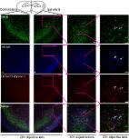
          Ischemic stroke is a clinically common cerebrovascular disease whose main risks include necrosis, apoptosis and cerebral infarction, all caused by cerebral ischemia and reperfusion (I/R). Ischemia and reperfusion-induced injury or apoptosis inhibition in human brain tissue may exert an irreplaceable protective effect on ischemic nerves. This process has particular significance for the treatment of stroke patients. However, the development of neuroprotective drugs remains challenging. Radix Scrophulariae, traditionally considered a valuable medicine, has been discovered to have neuroprotective effects. To explore the neuroprotective effects of an aqueous extract of Radix Scrophulariae (RSAE) on cerebral ischemia/reperfusion and their underlying mechanisms, oxygen-glucose deprivation and reperfusion (OGD/R)-induced PC12 cells were used, and a middle cerebral artery occlusion/reperfusion (MCAO/R) mouse model was established. In vitro results showed that 12.5 μg/mL RSAE markedly improved cell viability; inhibited LDH leakage; increased SOD, GSH-Px and CAT enzyme activity; stabilized the mitochondrial membrane potential; and reduced OGD-induced cell injury and apoptosis. Additionally, in vivo results preliminarily suggested that in MCAO/R model mice, RSAE treatments attenuated infarct volume; reduced brain water content and nitric oxide (NO) and malondialdehyde (MDA) concentrations; inhibited I/R-induced neurological deficits; reduced the levels of lactate dehydrogenase (LDH) leakage release; improved antioxidant capacity by upregulating SOD, GSH-Px and CAT enzyme activity; and reduced neuronal apoptosis, necrosis and loss of neurons. Moreover, it was found that RSAE upregulated the expression of Bcl-2 and downregulated the expression of Bax. In addition, the phosphorylation levels of MAPK signal pathways were elucidated via western blot analysis and immunohistochemical evaluation. In summary, this study investigated the neuroprotective effects and potential mechanisms of RSAE on focal cerebral I/R injury in mice. Radix Scrophulariae has been previously identified as a potential neuroprotective natural plant. Hence, our results may offer insight into discovering new active compounds or drugs for the treatment of ischemic stroke. Many new natural active chemicals in this extract may be discovered by chemical separation and identification and may provide new insights into therapeutic targets in stroke patients.

          Related collections

          Most cited references35

          • Record: found
          • Abstract: found
          • Article: found
          Is Open Access

          Attenuation of TNF-α-Induced Inflammatory Injury in Endothelial Cells by Ginsenoside Rb1 via Inhibiting NF-κB, JNK and p38 Signaling Pathways

          It is currently believed that inflammation plays a central role in the pathophysiology of atherosclerosis. Oxidative stress and redox-sensitive transcription factors are implicated in the process. Ginsenoside Rb1, a major active ingredient in processed Radix notoginseng, has attracted widespread attention because of its potential to improve cardiovascular function. However, the effects of ginsenoside Rb1 on tumor necrosis factor-α (TNF-α)-induced vascular endothelial cell injury and the underlying molecular mechanisms have never been studied. This study showed that TNF-α-induced oxidative stress, inflammation and apoptosis in human umbilical vein endothelial cells (HUVECs) could be attenuated by ginsenoside Rb1 pretreatment. Using JC-1, Annexin V/PI and TUNEL staining, and a caspase-3 activity assay, we found that Rb1 provided significant protection against TNF-α-induced cell death. Furthermore, Rb1 pretreatment could inhibit TNF-α-induced ROS and MDA production; increase the activities of SOD, CAT, and GSH-Px; and decrease the levels of IL-1β, IL-6, VCAM-1, ICAM-1, VEGF, MMP-2 and MMP-9. Importantly, the cytoprotective effects of Rb1 were correlated with NF-κB signaling pathway inhibition. Additionally, we found that Rb1 may suppress the NF-κB pathway through p-38 and JNK pathway activation, findings supported by the results of our experiments involving anisomycin (AM), a JNK and p38 activator. In conclusion, this study showed that ginsenoside Rb1 protects HUVECs from TNF-α-induced oxidative stress and inflammation by inhibiting JNK and p38. This inhibition suppressed NF-κB signaling and down-regulated the expression of inflammatory factors and apoptosis-related proteins.
            Bookmark
            • Record: found
            • Abstract: found
            • Article: found
            Is Open Access

            Neuroprotective effects of bilobalide on cerebral ischemia and reperfusion injury are associated with inhibition of pro-inflammatory mediator production and down-regulation of JNK1/2 and p38 MAPK activation

            Background Mitogen-activated protein kinase (MAPK) signaling pathways are implicated in inflammatory and apoptotic processes of cerebral ischemia and reperfusion (I/R) injury. Hence, MAPK pathways represent a promising therapeutic target. Exploring the full potential of inhibitors of MAPK pathways is a useful therapeutic strategy for ischemic stroke. Bilobalide, a predominant sesquiterpene trilactone constituent of Ginkgo biloba leaves, has been shown to exert powerful neuroprotective properties, which are closely related to both anti-inflammatory and anti-apoptotic pathways. We investigated the neuroprotective roles of bilobalide in the models of middle cerebral artery occlusion and reperfusion (MCAO/R) and oxygen-glucose deprivation and reoxygenation (OGD/R) of cerebral I/R injury. Moreover, we attempted to confirm the hypothesis that its protection effect is via modulation of pro-inflammatory mediators and MAPK pathways. Methods Male Sprague-Dawley rats were subjected to MCAO for 2 h followed by reperfusion for 24 h. Bilobalide was administered intraperitoneally 60 min before induction of middle cerebral artery occlusion (MCAO). After reperfusion, neurological deficit scores, infarct volume, infarct weight, and brain edema were assessed. Ischemic penumbrae of the cerebral cortex were harvested to determine superoxide dismutase (SOD), malondialdehyde (MDA), nitric oxide, TNF-α, interleukin 1β (IL-1β), p-ERK1/2, p-JNK1/2, and p-p38 MAPK concentration. Similarly, the influence of bilobalide on the expression of nitric oxide, TNF-α, IL-1β, p-ERK1/2, p-JNK1/2, and p-p38 MAPK was also observed in an OGD/R in vitro model of I/R injury. Results Pretreatment with bilobalide (5, 10 mg/kg) significantly decreased neurological deficit scores, infarct volume, infarct weight, brain edema, and concentrations of MDA, nitric oxide, TNF-α, IL-1β, and increased SOD activity. Furthermore, bilobalide (5, 10 mg/kg) pretreatment significantly down-regulated both p-JNK1/2 and p-p38 MAPK expression, whereas they had no effect on p-ERK1/2 expression in the ischemic penumbra. Supporting these observations in vivo, pretreatment with bilobalide (50, 100 μM) significantly down-regulated nitric oxide, TNF-α, IL-1β, p-JNK1/2, and p-p38 MAPK expression, but did not change p-ERK1/2 expression in rat cortical neurons after OGD/R injury. Conclusions These data indicate that the neuroprotective effects of bilobalide on cerebral I/R injury are associated with its inhibition of pro-inflammatory mediator production and down-regulation of JNK1/2 and p38 MAPK activation.
              Bookmark
              • Record: found
              • Abstract: found
              • Article: found
              Is Open Access

              TRPM7 inhibitor carvacrol protects brain from neonatal hypoxic-ischemic injury

              Background Our previous study found that suppression of TRPM7 reduced neuronal death in adult rat ischemic brain injury. It was reported that carvacrol blocked TRPM7 and attenuated brain injury in an adult rat MCAO model. The effects of carvacrol on neonatal stroke remain unknown. This study investigated the effects of carvacrol on neuronal injury and behavioral impairment after hypoxia-ischemia in neonatal mice and the potential signaling pathway underlying these effects. Results Carvacrol inhibited TRPM7 current in HEK293 cells over-expressing TRPM7 and TRPM7-like current in hippocampal neurons in a dose-dependent manner. Carvacrol (>200 μM) reduced OGD-induced neuronal injury in cortical neurons. 24 hours after HI, TRPM7 protein level in the ipsilateral hemisphere was significantly higher than in the contralateral hemisphere. Carvacrol (30 and 50 mg/kg) pre-treatment reduced brain infarct volume 24 hours after HI in a dose-dependent manner. Carvacrol pre-treatment also improved neurobehavioral outcomes. Furthermore, animals pre-treated with carvacrol had fewer TUNEL-positive cells in the brain compared to vehicle-treated animals 3 days after HI. Carvacrol pre-treatment also increased Bcl-2/Bax and p-Akt/t-Akt protein ratios and decreased cleaved caspase-3 protein expression 24 hours after HI. Conclusions Carvacrol pre-treatment protects against neonatal hypoxic-ischemic brain injury by reducing brain infarct volume, promoting pro-survival signaling and inhibiting pro-apoptotic signaling, as well as improving behavioral outcomes. The neuroprotective effect may be mediated by the inhibition of TRPM7 channel function. Carvacrol is a potential drug development target for the treatment of neonatal stroke.
                Bookmark

                Author and article information

                Journal
                Molecules
                Molecules
                molecules
                Molecules : A Journal of Synthetic Chemistry and Natural Product Chemistry
                MDPI
                1420-3049
                19 September 2018
                September 2018
                : 23
                : 9
                : 2401
                Affiliations
                [1 ]Beijing Key Laboratory of Innovative Drug Discovery of Traditional Chinese Medicine (Natural Medicine) and Translational Medicine, Institute of Medicinal Plant Development, Peking Union Medical College and Chinese Academy of Medical Sciences, Beijing 100193, China; xbmeng@ 123456implad.ac.cn (X.M.); ginseng123@ 123456163.com (W.X.); shengjupan@ 123456163.com (Q.X.); liantianparper@ 123456sina.com (T.L.)
                [2 ]Key Laboratory of Bioactive Substances and Resource Utilization of Chinese Herbal Medicine, Ministry of Education, Beijing 100193, China
                [3 ]Key Laboratory of Efficacy Evaluation of Chinese Medicine against Glycolipid Metabolic Disorders, State Administration of Traditional Chinese Medicine, Beijing 100193, China
                [4 ]Zhongguancun Open Laboratory of the Research and Development of Natural Medicine and Health Products, Beijing 100193, China
                Author notes
                [* ]Correspondence: wym91116@ 123456163.com (X.X.); sunguibopaper@ 123456163.com (G.S.); sunxiaobopaper@ 123456163.com (X.S.); Tel.: +86-10-5783-3220 (G.S.); +86-10-5783-3013 (X.S.)
                [†]

                These authors contributed equally to this paper.

                Author information
                https://orcid.org/0000-0002-5062-7843
                Article
                molecules-23-02401
                10.3390/molecules23092401
                6225418
                30235876
                fddcd492-abb9-4575-be4d-a2e39f0099d9
                © 2018 by the authors.

                Licensee MDPI, Basel, Switzerland. This article is an open access article distributed under the terms and conditions of the Creative Commons Attribution (CC BY) license ( http://creativecommons.org/licenses/by/4.0/).

                History
                : 19 August 2018
                : 13 September 2018
                Categories
                Article

                radix scrophulariae,cerebral ischemia stroke,middle cerebral artery occlusion-reperfusion,apoptosis,oxygen-glucose deprivation and reperfusion,mapk pathways

                Comments

                Comment on this article