293
views
0
recommends
+1 Recommend
1 collections
    0
    shares
      scite_
       
      • Record: found
      • Abstract: found
      • Article: found
      Is Open Access

      Resolution of complex ends by Nonhomologous end joining - better to be lucky than good?

      review-article
      Bookmark

            Abstract

            The Nonhomologous end joining pathway is essential for efficient repair of chromosome double strand breaks. This pathway consequently plays a key role in cellular resistance to break-inducing exogenous agents, as well as in the developmentally-programmed recombinations that are required for adaptive immunity. Chromosome breaks often have complex or “dirty” end structures that can interfere with the critical ligation step in this pathway; we review here how Nonhomologous end joining resolves such breaks.

            Main article text

            Double strand break repair and complex end structures

            DNA double strand breaks (DSBs) arise after replication, aberrant repair of spontaneous damage, and exposure to exogenous damaging agents, especially those used in cancer therapies. DSBs are also intermediates in several developmentally-programmed recombinations. Failed DSB repair is typically lethal, while aberrant DSB repair can lead to developmental defects, progeria, and cancer. Repair pathways include Homologous recombination (HR), Nonhomologous end joining (NHEJ), and Alternate end joining (Alt-EJ) (reviewed in e.g. [1]). Importantly, HR is dependent on extensive (100s to 1000s of nucleotides) DNA synthesis, a sister chromatid template to direct this synthesis, and a homology search step needed to find the template in a sister chromatid. In contrast, NHEJ is primarily a ligation reaction and can act independently of S-phase restricted sister chromatids, dNTP generation [2], and other requirements for extensive synthesis. Finally, a fraction of ligation-mediated repair is independent of factors required for classically defined NHEJ, and is thus termed “Alt-EJ”.

            The primary disadvantage to resolving ends by ligation is that biological sources of DSBs often produce “dirty” or complex end structures that can interfere with this step (Figure 1). DNA flanking the break may possess nucleotide damage, most frequently oxidized bases, various classes of abasic sites, and 3 phosphate or 5 hydroxyl termini [3]. Such damage is especially likely in the case of ionizing radiation-induced breaks, which are associated with damage clusters [3,4]. DSB ends may be further occluded by proteins, both non-covalently associated (e.g. chromatin) and covalently adducted type II topoisomerases [5]. Ends can also possess secondary structures including hairpins or, after a pair of ends are aligned together, gaps, mismatches, or flaps.

            Figure 1

            Biological sources of DSBs generate complex end structures. Examples include oxidized nucleotides, protein occlusions, and secondary structures.

            NHEJ resolves complex ends by employing a sophisticated machine engineered to facilitate ligation despite ligation-blocking lesions. A series of core factors are necessary and sufficient to recognize ends, align a pair of ends together, and perform the ligation step (Figure 2). Core factors include 1) Ku; a DNA end binding heterodimer of 70 and 80 kD subunits (Ku70, Ku80), 2) XRCC4-ligase IV (X4-LIV); an obligate oligomer of a ligase catalytic subunit (DNA ligase IV) and scaffolding subunit (XRCC4), 3) XRCC4-like factor (XLF, also termed Cernunnos), and 4) DNA dependent protein kinase catalytic subunit (DNA-PKcs); a 450 kD kinase recruited to ends by Ku (reviewed in [6]). However, additional factors are required, both to integrate NHEJ with the DNA damage response and local chromatin structure [7], as well as (to be discussed here) to help this core machine resolve complex ends (Table 1). NHEJ employs these additional factors according to strategies (Figure 2) for resolving complex ends we suggest can be roughly categorized as 1) tolerance (Figure 3), 2) end “cleaning” (Figure 4), and 3) trial-and-error (Figure 5).

            Figure 2

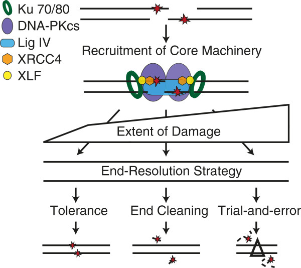
            NHEJ end-resolution strategies. Resolution of complex ends by NHEJ first requires the recruitment of the core machinery (Ku, DNA-PKcs, Ligase IV, XRCC4, and XLF). The type and extent of damage varies, and this in turn probably dictates choice of strategy.

            Table 1

            End processing factors

            Factor*Activity
            APTX
            Removes 5-adenylate adducts [40]
            PNKP
            Removes 3 phosphates and phosphorylates 5 hydroxyls [45]
            APLF
            Histone chaperone [59] 3-5 exonuclease, endonuclease [56,61]
            TDP1
            Removes Top I adducts [63], 3 deoxyribose fragments [47,67,68]
            TDP2
            Removes Top II adducts [64]
            XRCC5,XRCC6 (Ku)
            Removes 5-dRP residues and abasic sites [27]
            POLM (Pol λ)
            Fills in gaps when ends align with no complementarity [90]
            POLL (Pol μ)
            Fills in gaps when ends are partly complementary [86,90]
            DCLRE1C (Artemis)
            Endonuclease, 5-3 exonuclease [100]
            WRN
            3-5 exonuclease [121,122] and 3-5 helicase [120]
            MRE11/RAD50/NBN (MRN)
            3-5 exonuclease, endonuclease [101,129]
            SETMAR (Metnase)Endonuclease/exonuclease [103]

            *HUGO gene nomenclature: APTX, aprataxin; PNKP, polynucleotide kinase 3-phosphatase; APLF, aprataxin and PNKP like factor; TDP1, tyrosyl-DNA phosphodiesterase 1; TDP2, tyrosyl-DNA phosphodiesterase 2; XRCC5,XRCC6 (Ku80, Ku70), X-ray repair complementing defective repair in Chinese hamster cells 5/6; POLM, polymerase mu; POLL, polymerase lambda; DCLRE1C (Artemis), DNA cross-link repair 1C; WRN, Werner syndrome; MRE11/RAD50/NBN, meiotic recombination 11 homolog/RAD50 homolog/nibrin (Nbs1), SETMAR, SET domain and mariner transposase fusion gene (Metnase).

            Figure 3

            Joining by tolerance. The NHEJ core machinery can be associated with an XLF-XRCC4 filament that may stabilize aligned complex end structures (i) sufficiently to support transfer of the adenylate from the ligase to 5phosphate terminus (ii), possibly through to complete ligation (iii). If ligation aborts, the 5 adenylated (AMP) intermediate can be removed by Aprataxin (APTX) (iv). DNA-PKcs-directed phosphorylation of XLF and XRCC4 can disrupt the XLF-XRCC4 filament (v) to allow processing factors access to the DNA to remove the obstruction.

            Figure 4

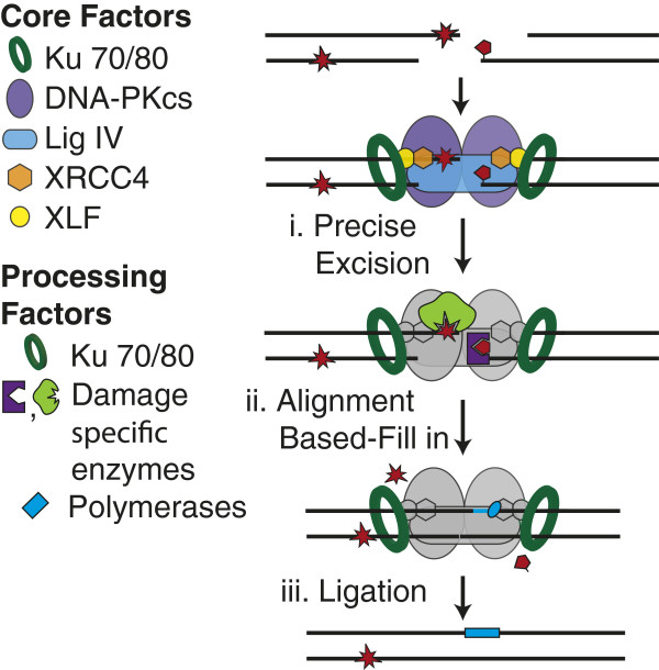
            End cleaning. Ligation-blocking damage can be excised by enzymes (e.g. APTX, PNKP, APLF, Tdps, Ku; purple and green proteins) that recognize and remove specific end structures (i). Family X polymerases (light blue oval) can replace the excised DNA (ii) prior to ligation (iii).

            Figure 5

            Trial and error. Ends can go through sequential rounds of end alignment (i) and processing (ii) (primarily nucleases; black Pac-man) until ligated. Alternatively, ends can be resected (iii) and Ku released, enabling resolution of ends by homologous recombination (HR) or alternative end-joining (Alt-EJ) pathways.

            Tolerance of complex ends by low fidelity ligation

            Ligation initiates with the transfer of an adenosine monophosphate from a lysine within the ligase active site to the 5 phosphate (Figure 3, step ii). The final step in ligation has similarities to DNA synthesis, as the new phosphodiester bond is made by a phosphoryl transfer reaction, where the 3 hydroxyl terminus of one strand performs a nucleophillic attack on the activated 5 phosphate terminus of the second (Figure 3, step iii) (reviewed in [8]). Importantly, most DNA ligases resemble polymerases in that they are most active when joining strands with termini complementary to a “template” strand. Thus, in the same manner as polymerases, ligases can be considered as high or low fidelity according to the degree to which they tolerate mispairs or other helix distortions in DNA flanking the bond to be made.

            The XLF-X4-LIV splint and low fidelity ligation

            X4-LIV can join together strand termini with flanking mispairs more readily than other ligases [9,10], and thus can be considered to be of low fidelity. Importantly, XLF specifically promotes low-fidelity ligation [11,12]. Dimers of XLF interact with dimers of XRCC4 and DNA [13-18], resulting in DNA- bound (XLF-XRCC4)N filaments [14,19-23] that eventually include or terminate in a X4-LIV complex (Figure 3, step i). These filaments are thought to act as a protein-“splint”, stabilizing an aligned pair of ends. The splint could account for reduced ligation fidelity by suppressing helix distortions associated after alignment of complex end structures, or by simply increasing the time the active site has to work with end alignments with poorly oriented termini. Low-fidelity ligation is advantageous for NHEJ as it increases the spectrum of complex ends that can be ligated together directly without prior end processing.

            However, there is a limit to what NHEJ’s ligase can tolerate [9,24-30], even with the XLF-X4-LIV splint. Notably, DNA-PKcs-mediated phosphorylation of multiple sites within XLF and XRCC4 disrupts the filament (Figure 3, step v) [31]. This may relax (or release) unproductive complexes of the ligase and aligned ends to give better access to end processing enzymes required for other NHEJ strategies (e.g, Figures 4, 5).

            Additionally, like damage tolerance by translesion polymerases in replication, tolerance of complex end structures by NHEJ is a form of procrastination – mismatched and damaged nucleotides flanking the break site are not repaired and thus retained in the joined product (junction). Unresolved damage will interfere with subsequent transcription or replication through the junction. Additionally, attempted repair by other pathways (e.g. base excision repair; BER) of residual damage clusters in junctions risks re-breaking the site [32-36]. Sustained localization of NHEJ factors (e.g. Ku, X4-LIV, XLF) after joining may even help regulate BER activity.

            Damage-specific end cleaning

            Ligases cannot join ends unless they have 5 phosphate and 3 hydroxyl termini, regardless of how stably a pair of aligned termini are juxtaposed. In addition, NHEJ’s ligation step is blocked by terminal or near-terminal abasic sites [24-27,29] and when lesions or mismatches in end structures are sufficiently helix distorting. Common terminus-blocking lesions can be excised and the potentially resulting gaps filled in before ligation (Figure 4). As with other repair pathways (e.g. BER), NHEJ can thus fully restore sequence at DSB sites, even when the break was associated with ligation blocking damage [24].

            To this end, NHEJ employs an array of enzymes that partly overlap with BER and single strand break repair (SSBR). Indeed, three of these factors – aprataxin (APTX), polynucleotide kinase/phosphatase (PNKP), and aprataxin and polynucleotide kinase/phosphatase like factor (APLF) - employ N-terminal forkhead associated (FHA) domains to mediate their participation in both NHEJ and BER/SSBR pathways (reviewed in [37]). These domains physically interact with XRCC4 [38] and XRCC1 [39] to direct their participation in NHEJ and BER/SSBR, respectively. FHA domain-mediated interactions are stimulated by phosphorylation of XRCC4 and XRCC1 by casein kinase II.

            Aprataxin

            As noted above, there is a limit to what the ligase can tolerate. Attempts to tolerate complex end structures can result in ligation failure at an intermediate step, after adenylation of the 5 terminus but before the final phosphoryl transfer (Figure 3, step ii). A new ligase IV molecule cannot act on the 5 adenylated product of aborted ligation; Aprataxin (APTX) resets the substrate for another ligation attempt by removing 5-adenylate adducts (Figure 3, step iv), as mediated by APTX’s zinc-finger-histidine triad (HIT) domain [40].

            Mutations in APTX account for several neurodegenerative disorders, including ataxia with oculomotor apraxia type 1 (AOA1) [41,42]. However, sensitivity of APTX deficient cells to various DNA damaging agents is mild [43], and it has been difficult to detect measurable differences in either SSBR or DSBR ([44] and references therein). APTX is thus argued to act on a minor subset of breaks [44], with the consequences of failed action in an organism possibly disproportionate to the low frequency of these events.

            Polynucleotide kinase/phosphatase

            5 hydroxyl and 3 phosphate termini are generated directly by reactive oxygen species, after metabolization of certain strand breaks (3- phosphoglycolate, 3-phosphotyrosine), or by the action of endo-VIII-like glycosylases (Neil1 or Neil2). PNKP has two catalytic domains sufficient to prepare such termini for ligation: a central domain that removes 3 phosphates, and a C-terminal domain that phosphorylates 5 hydroxyls [45] (reviewed in [46]). PNKP was also shown to act coordinately with TDP1 to remove the 3 phosphate generated after removal of 3-phosphoglycolate residues [47].

            Mutations in PNKP result in microcephaly with early onset intractable seizures and developmental delay (MCSZ) [48]. A role for PNKP specifically in double strand break repair is supported by sensitivity of PNKP deficient cells to ionizing radiation [48,49], and PNKP is required for NHEJ of ends without 5 phosphate in cell extracts [50]. PNKP’s contribution to radiation sensitivity relies to some extent on damage-dependent phosphorylation of sites in PNKP by DNA-PKcs and ATM [51,52].

            Aprataxin and polynucleotide kinase/phosphatase like factor

            APLF (also referred to as PALF, C2orf13, and Xip1) possesses a tandem pair of poly(ADP-ribose) binding zinc finger (PBZ) motifs [53], which mediate recruitment of APLF to damage [54-57] after poly(ADP) ribose polymerase-3 (PARP-3) modification of flanking chromatin [58]. Disruption of APLF’s PBZ domains attenuates X4-LIV accumulation at DSB ends in cells, which in turn results in defects in NHEJ of radiation induced breaks and DSB intermediates in class switch recombination [58]. This may be at least partly because a network of interactions between APLF, Ku [56,57], X4-LIV [57,58], DNA, and poly(ADP-ribose) chromatin [57,58] could be required for stable assembly of an NHEJ complex at ends. APLF also possesses a conserved C-terminal domain with a NAP1L family histone chaperone motif, which is sufficient to promote assembly and disassembly of nucleosomes and nucleosome substructures in vitro[59]. Ku can recognize DSB ends even when on the surface of a nucleosome [28], but an active NHEJ complex, including X4-LIV and DNA-PKcs, requires at least 60 bp of free DNA flanking the end [60] (probably more with an (XLF-X4)N splint). APLF, perhaps triggered by coordinate recognition of ends by Ku and PBZ mediated interactions with flanking poly(ADP-ribose) modified chromatin, could direct a very limited remodeling of nucleosomes flanking broken ends to make room for subsequent loading of X4-LIV. APLF may additionally act as an exonuclease and structure-specific endonuclease [56,61] to resolve mismatches and flaps.

            Tyrosyl DNA phosphodiesterases

            Topoisomerases (Top I, Top II) resolve DNA topological stress associated with replication and transcription. They employ a cleavage/ligation cycle with an intermediate step where a tyrosine in the topoisomerase is covalently linked to a strand break through 3 or 5 phosphate termini (Top I and top II, respectively) [62]. The cleavage/ligation equilibrium can be altered (e.g. after treatment with topoisomerase poisons) such that cells accumulate strand breaks with termini adducted to a topoisomerase through their active site tyrosine [5]. If a type I topoisomerase aborts, the topoisomerase is adducted to a single strand break, 3 phosphate terminus; if a type II topoisomerase aborts, the topoisomerase is adducted to DSB 5 phosphate termini. Adducted topoisomerases can be removed from DNA strand termini by reversal of the covalent intermediate, through the action of tyrosyl-DNA phosphodiesterases Tdp1 [63] and Tdp2 [64].

            Tdp1’s primary substrates are the 3-phosphotyrosine adducts generated by aborted type I topoisomerase activity, either at SSBs, or DSBs generated after replication through unrepaired SSBs [65,66]. Epistasis analysis indicates Tdp1 activity on even the DSB-associated products is upstream of repair by HR in S. cerevisae[66]. However, Tdp1 additionally has activity on other 3 phosphate adducts including 3-phosphoglycolates [67-71] (Table 1), the most common class of nucleotide damage associated with ionizing radiation induced strand breaks. Tdp1 is required for resolution by NHEJ of substrates with 3-phosphoglycolate termini in a cell extract model [47], and patients with Tdp1 mutations (Spinocerebellar ataxia with axonal neuropathy 1; SCAN-1) [72,73] are sensitive to radiomimetic drugs that can introduce strand breaks with 3-phosphoglycolate termini [47,74]. At the same time there are significant backup pathways active in cells [75], possibly explaining why SCAN1 cells are neither severely sensitive to ionizing radiation [71], nor possess obvious defects in rates of DSB repair after ionizing radiation [76].

            Tdp2 (also termed TTRAP; TRAF and TNF receptor-associated protein, and EAPII; ETS1 associated protein II) is most active in removing tyrosines adducted to 5 phosphates at DSB ends [64,77], a product of aborted type II topoisomerase action. Consistent with this specificity, Tdp2 is essential for resistance of chicken DT-40 cells to type II topoisomerase poisons (e.g. etoposide) [78]. Tdp2 could in principle participate in either HR or NHEJ pathways for DSB repair, and there are as yet no reported epistasis analyses or physical interactions specifically linking Tdp2 to either. Nevertheless, a role for Tdp2 uniquely within NHEJ seems most likely. Unlike Top I adducts, there is little use for “clean” removal of Top II adducts within the HR pathway, since the 5 strand must anyway be extensively resected as a pre-requisite for the homology search step. Indeed, removal of the Top II-like Spo11 adduct at DSB intermediates during meiotic HR relies on the Mre11/Nbs1/CtIP complex; this latter pathway is apparently Tdp2 independent, as the excised Spo11 is still adducted to a short oligonucleotide [79].

            Ku

            DSB with associated abasic sites, either 5 terminal (5-deoxyribose phosphate; 5-dRP) or near-terminal (apurinic/apyrimidinic; AP), can be generated directly by ionizing radiation. However, they are probably more frequently associated with DSB products of aborted base excision repair, including the DSB intermediates in immunoglobulin class switch recombination [80,81]. Regardless of source, NHEJ cannot join such ends together both in vitro (whether or not XLF is present) [25-27,29] or in cells [27,29] unless the abasic site is excised. Excision of these abasic sites is mediated both in vitro and in cells primarily by the Ku heterodimer which, in addition to its primary role in recognizing ends and recruiting other factors, is a 5-dRP/AP lyase [27]. Notably, Ku’s 5-dRP/AP activity is primarily restricted to substrates where incision is both necessary and sufficient to prepare ends for the ligation step [29]. Specifically, Ku is much less active on abasic sites near 3 termini, where incision by a lyase would leave a ligation blocking 3-α, β-unsaturated aldehyde. Ku is similarly much less active when abasic sites near 5 termini are significantly embedded in duplex DNA, a context where the abasic site no longer blocks the ligation step. The latter substrate specificity is essentially non-overlapping relative to abasic site metabolizing enzymes implicated in BER (AP endonuclease, pol β), whose activities are mostly restricted to sites with significant (>4 bp) flanking dsDNA [26,27,29].

            Family X polymerases

            A variety of polymerases have been implicated in NHEJ (also reviewed in [82]), but the majority of evidence favors a primary role for several members of the mammalian family X polymerase: Pol λ, Pol μ, and Terminal deoxynucleotidyl transferase (TdT). All three polymerases possess homologous N-terminal BRCT (Breast cancer associated carboxy-terminal) domains [83] that promote formation of a complex including the polymerase, Ku and X4-LIV at DNA ends [84-90]. BRCT domains have no impact on intrinsic catalytic activity but are essential for the participation of the polymerases in NHEJ, emphasizing the importance of coupling their catalytic activities to a complex of aligned ends [85,87,90]. The three polymerases have distinct substrate requirements and activities, with decreasing dependence on template strand, in order Pol λ>Pol μ>TdT [90]. All three have been clearly implicated in repair by NHEJ of intermediates in V(D)J recombination [91-94], with TdT’s entirely template independent activity observed only during V(D)J recombination by virtue of its restricted expression [95]. The other two polymerases are expressed in all cell types and have overlapping activities [83,96], making it difficult to parse their relative contributions. In general, they fill in gaps present after alignment of broken ends (the gap typically a consequence of prior excision of damaged nucleotides by enzymes discussed above). The action of Pol μ and Pol λ thus further extends parallels between NHEJ and BER/SSBR, and indeed Pol λ has roles in both [97-99], similar to APTX, PNKP and APLF.

            Trial and error

            NHEJ can use enzymes that specifically and precisely resolve many of the end structures expected to interfere with ligation of DSB ends. This means some fraction of NHEJ can proceed by an ordered, three-step strategy (excision, replacement, ligation; Figure 4) essentially equivalent to that used by base excision repair. However, such a strategy will not always suffice for DSBs. Some end structures do not appear to be readily resolved by the available enzymes (e.g. reduced abasic sites; [27,29]). Additionally, it is not yet clear how well NHEJ can identify the enzyme appropriate to a given context; this may be particularly challenging at ends associated with densely-clustered damage. A fraction of NHEJ could thus proceed by “trial and error”. In this strategy, ends would be subject to sequential ligation attempts (Figure 5, step i, “trial”) and end processing (Figure 5, step ii, “error”), with these steps repeated until a substrate for ligation is generated. We suggest that efficient resolution by this strategy implies a need for regulated transition between steps.

            This strategy employs processing factors that are more likely to be endonucleases that target secondary structures (single stranded overhangs, flaps, hairpins) and exonucleases, including Artemis [100], MRN [101], Werner’s syndrome protein [102], APLF [61], and Metnase [103]. Substrate specificities of these nucleases can be overlapping, and are generally less precisely targeted than the damage specific activities discussed above. Importantly, the latter characteristic allows them to aid in resolution of a wider variety of blocking lesions (more flexible), but the ensuing products are typically associated with greater deletion of DNA flanking the break site and are more heterogeneous. Initial overhang sequence complementarity will thus be reduced or lost, necessitating additional rounds of processing for ligation.

            Artemis

            Artemis has been associated with 5>3 exonuclease activity, but can be primarily linked to NHEJ through an important role for its structure-specific endonuclease activity [100]. The latter is mediated by a metallo β lactamase associated with CPSF Artemis SNM1/PSO2 domain [104-106]. Nuclease activity is dependent on the additional presence of DNA-PKcs at ends [100], as well as DNA-PKcs autophosphorylation [107]. Artemis’s C terminus is also phosphorylated by both DNA-PKcs and the related Ataxia Telangiectasia Mutated (ATM) kinase [106,108-112], and this may further help regulate Artemis activity. Most notably, Artemis is required for opening hairpins at broken ends. Such structures are critical intermediates in the assembly of antigen specific receptors by V(D)J recombination, consequently loss of Artemis function results in severe immunodeficiency [100,113]. Loss of Artemis also confers cellular sensitivity to IR [113-115], suggestive of a role for this nuclease in resolving complex end structures expected from IR-induced breaks. Consistent with this, Artemis generally cleaves at ssDNA/dsDNA transitions, and thus can remove extended (>4 nucleotide) ssDNA overhangs and flaps [100,109], as well as overhangs with ligation blocking nucleotide damage [116]. Notably, products of Artemis nuclease activity are heterogenous, with sites of cleavage often distributed over a 3-6 nucleotide range [100,109,116,117]. For example, Artemis, like Tdp1, can excise 3-phosphoglycolate termini in vitro but Artemis typically deletes a variable number of nucleotides in addition to the 3-phosphoglycolate residue [116]. Both the increased deletion and deletion heterogeneity associated with Artemis activity will more frequently necessitate additional rounds of processing before ends can be ligated, relative to the damage specific activities described in the previous section [87].

            Werners syndrome protein

            Werners syndrome is associated with progeria [118] and mild cellular sensitivity to ionizing radiation [119]. The Werners syndrome gene product (WRN) has both 3>5 exonuclease and helicase activity [120-122], and associates with NHEJ core factors Ku [102,123] and X4-LIV [124]. WRN activity is further regulated by DNA-PKcs [119,125], and can cooperate with these NHEJ factors to promote ligation in vitro after degradation of non-complementary overhangs [124]. Notably, WRN can degrade through oxidative damage in the presence of Ku in some contexts [126,127], possibly promoting NHEJ at ends where the density of break-associated damage is too high for damage specific resolutions.

            Mre11/Rad50/Nbs1

            The Mre11/Rad50/Nbs1 (MRN) complex (or Mre11/Rad50/Xrs2 in S. cerevisae) is required for efficient sensing of double strand breaks and helps bridge broken ends together (reviewed in [128]). Similar to Artemis, MRN also has 3>5 exonuclease and single strand specific endonuclease activities (including hairpin opening activity) [101,129], but while MRN has an important role in mammalian NHEJ [130-132] this role is only partly reliant on MRN’s nuclease activity [132]. Notably, MRN nuclease activity is essential for removal of Spo11 covalently adducted to 5 termini (a substrate analogous to aborted Topoisomerase II complexes) [79,133]. However, activity of the MRN and CtIP complex on this substrate precedes a more processive resection of 5 ends (reviewed in [134]) that primarily channels these breaks to repair by the homologous recombination pathway. Indeed, MRN/CtIP may perform this function whenever end structures cannot be resolved by any of the NHEJ strategies discussed (Figure 5 step iii), allowing these ends to be resolved instead by either alternate end joining or homologous recombination. Consistent with this idea, MRN’s nuclease activity has been implicated in release of Ku from DNA ends [135], thereby precluding further futile NHEJ attempts.

            Concluding remarks: is it better to be lucky than good?

            Complex end structures are diverse, explaining why NHEJ employs three distinguishable strategies (Figure 2) and a host of different processing activities (Table 1). Moreover, processing activities have varying degrees of substrate specificity, allowing NHEJ to balance precision with flexibility during end processing. When processing is required, these steps are appropriately ordered and coupled to the ligation step within a multiprotein machine. NHEJ is therefore indispensible for efficient resolution of complex end structures.

            Ideally, NHEJ chooses a resolution strategy and processing factor in a manner that optimizes the efficiency and fidelity of product. With respect to strategy, there may be a means for sensing the extent of damage first and specifically choosing the appropriate strategy (Figure 2). Alternatively, strategies may be employed hierarchically, starting with damage tolerance, followed by damage specific end cleaning, followed by trial and error. Additionally, the choice of processing factor may be determined only by its affinity for a specific substrate, or might additionally be regulated by access to its substrate.

            Accumulating evidence implicates DNA-PKcs kinase activity as the primary factor that could determine both choice of strategy and processing factor. DNA-PKcs kinase activity is dependent on end context, both in terms of whether a pair of ends can be aligned together [136,137], but also as a reflection of differences in end structure [138-140]. Additionally (as noted above), DNA-PKcs-mediated phosphorylation of Artemis [107-112], PNKP [51,52], Tdp1 [65], WRN [119,125], and XLF-X4-LIV [31] can affect the activity of these proteins. However, DNA-PKcs itself is probably the most relevant target (i.e., autophosphorylation), as there are in excess of 30 different sites (reviewed in [141]) that together may be sufficient for a “code,” where phosphorylation of different patches has distinguishable effects on end access [142-144].

            Events in the resolution of complex ends by NHEJ can thus be, to some extent, left to chance, but can also be precisely scripted. So, is it better to be lucky than good? Why not both?

            Competing interests

            The authors declare no competing interests.

            Authors’ contributions

            NTS, CAW, and DAR wrote the manuscript, and N.T.S. assembled the figures. All authors read and approved the final manuscript.

            Acknowledgements

            We thank the members of the Ramsden laboratory for their helpful discussion. This work was supported by NIH NCI grants CA 84442 and CA 97096 to D.A.R.

            References

            1. HiomKCoping with DNA double strand breaksDNA Repair2010912561263[PubMed]

            2. BurkhalterMDRobertsSAHavenerJMRamsdenDAActivity of ribonucleotide reductase helps determine how cells repair DNA double strand breaksDNA Repair2009812581263[PubMed]

            3. WardJFThe complexity of DNA damage: relevance to biological consequencesInt J Radiat Biol199466427432[PubMed]

            4. GoodheadDTInitial events in the cellular effects of ionizing radiations: clustered damage in DNAInt J Radiat Biol199465717[PubMed]

            5. NitissJLTargeting DNA topoisomerase II in cancer chemotherapyNat Rev Cancer20099338350[PubMed]

            6. LieberMRThe mechanism of double-strand DNA break repair by the nonhomologous DNA end-joining pathwayAnnu Rev Biochem201079181211[PubMed]

            7. LukasJLukasCBartekJMore than just a focus: the chromatin response to DNA damage and its role in genome integrity maintenanceNat Cell Biol20111311611169[PubMed]

            8. EllenbergerTTomkinsonAEEukaryotic DNA ligases: structural and functional insightsAnnu Rev Biochem200877313338[PubMed]

            9. GuJLuHTippinBShimazakiNGoodmanMFLieberMRXRCC4:DNA ligase IV can ligate incompatible DNA ends and can ligate across gapsEMBO J200726114[PubMed]

            10. WangYLamarcheBJTsaiMDHuman DNA ligase IV and the ligase IV/XRCC4 complex: analysis of nick ligation fidelityBiochemistry20074649624976[PubMed]

            11. GuJLuHTsaiAGSchwarzKLieberMRSingle-stranded DNA ligation and XLF-stimulated incompatible DNA end ligation by the XRCC4-DNA ligase IV complex: influence of terminal DNA sequenceNucleic Acids Res20073557555762[PubMed]

            12. TsaiCJKimSAChuGCernunnos/XLF promotes the ligation of mismatched and noncohesive DNA endsProc Natl Acad Sci USA200710478517856[PubMed]

            13. AhnesorgPSmithPJacksonSPXLF interacts with the XRCC4-DNA ligase IV complex to promote DNA nonhomologous end-joiningCell2006124301313[PubMed]

            14. AndresSNModestiMTsaiCJChuGJunopMSCrystal structure of human XLF: a twist in nonhomologous DNA end-joiningMol Cell20072810931101[PubMed]

            15. HentgesPPAhnesorgPPPitcherRSRBruceCKCKyselaBBGreenAJABianchiJJWilsonTETJacksonSPSDohertyAJAEvolutionary and functional conservation of the DNA non-homologous end-joining protein, XLF/CernunnosJ Biol Chem20062813751737526[PubMed]

            16. LiYChirgadzeDYBolanos-GarciaVMSibandaBLDaviesORAhnesorgPJacksonSPBlundellTLCrystal structure of human XLF/Cernunnos reveals unexpected differences from XRCC4 with implications for NHEJEMBO J200727290300[PubMed]

            17. LuHPannickeUSchwarzKLieberMRLength-dependent binding of human XLF to DNA and stimulation of XRCC4.DNA ligase IV activityJ Biol Chem20072821115511162[PubMed]

            18. SulekMYarringtonRMcGibbonGBoekeJDJunopMA critical role for the C-terminus of Nej1 protein in Lif1p association, DNA binding and non-homologous end-joiningDNA Repair2007618051818[PubMed]

            19. AndresSNVergnesARisticDWymanCModestiMJunopMA human XRCC4-XLF complex bridges DNANucleic Acids Res20124018681878[PubMed]

            20. HammelMReyMYuYManiRSClassenSLiuMPiqueMEFangSMahaneyBLWeinfeldMXRCC4 protein interactions with XRCC4-like factor (XLF) create an extended grooved scaffold for DNA ligation and double strand break repairJ Biol Chem20112863263832650[PubMed]

            21. HammelMYuYFangSLees-MillerSPTainerJAXLF regulates filament architecture of the XRCC4· ligase IV complexStructure/Folding and Design20101814311442

            22. OchiTWuQChirgadzeDYGrossmannJGBolanos-GarciaVMBlundellTLStructural insights into the role of domain flexibility in human DNA ligase IVStructure20122012121222[PubMed]

            23. RoparsVDrevetPLegrandPBaconnaisSAmramJFaureGMárquezJAPiétrementOGueroisRCallebautIStructural characterization of filaments formed by human Xrcc4–Cernunnos/XLF complex involved in nonhomologous DNA end-joiningProc Natl Acad Sci USA20111081266312668[PubMed]

            24. ChenSInamdarKVPfeifferPFeldmannEHannahMFYuYLeeJWZhouTLees-MillerSPPovirkLFAccurate in vitro end joining of a DNA double strand break with partially cohesive 3′-overhangs and 3′-phosphoglycolate termini.J Biol20012762432324330

            25. DattaKPurkayasthaSNeumannRDPastwaEWintersTABase damage immediately upstream from double-strand break ends is a more severe impediment to nonhomologous end joining than blocked 3′-termini.Radiat Res201017597112[PubMed]

            26. DobbsTAPalmerPManiouZLomaxMEO’NeillPInterplay of two major repair pathways in the processing of complex double-strand DNA breaksDNA Repair2008713721383[PubMed]

            27. RobertsSAStrandeNBurkhalterMDStromCHavenerJMHastyPRamsdenDAKu is a 5′-dRP/AP lyase that excises nucleotide damage near broken ends.Nature201046412141217[PubMed]

            28. RobertsSASRamsdenDADLoading of the nonhomologous end joining factor, Ku, on protein-occluded DNA endsJ Biol Chem20072821060510613[PubMed]

            29. StrandeNRobertsSAOhSHendricksonEARamsdenDASpecificity of the dRP/AP lyase of Ku promotes nonhomologous end joining (NHEJ) fidelity at damaged endsJ Biol Chem20122871368613693[PubMed]

            30. SearsCRTurchiJJComplex cisplatin-double strand break (DSB) lesions directly impair cellular non-homologous end-joining (NHEJ) independent of downstream damage response (DDR) pathwaysJ Biol Chem20122872426324272[PubMed]

            31. RoySAndresSNVergnesANealJAXuYYuYLees-MillerSPJunopMModestiMMeekKXRCC4’s interaction with XLF is required for coding (but not signal) end joiningNucleic Acids Res20124016841694[PubMed]

            32. GulstonMProcessing of clustered DNA damage generates additional double-strand breaks in mammalian cells post-irradiationNucleic Acids Res20043216021609[PubMed]

            33. MaWWestmorelandJWGordeninDAResnickMAAlkylation base damage is converted into repairable double-strand breaks and complex intermediates in G2 cells lacking AP endonucleasePLoS Genet20117e1002059[PubMed]

            34. MalyarchukSCastoreRHarrisonLDNA repair of clustered lesions in mammalian cells: involvement of non-homologous end-joiningNucleic Acids Res20083648724882[PubMed]

            35. MalyarchukSCastoreRHarrisonLApex1 can cleave complex clustered DNA lesions in cellsDNA Repair2009813431354[PubMed]

            36. YangNGalickHWallaceSSAttempted base excision repair of ionizing radiation damage in human lymphoblastoid cells produces lethal and mutagenic double strand breaksDNA Repair2004313231334[PubMed]

            37. CaldecottKWSingle-strand break repair and genetic diseaseNat Rev Genet20089619631[PubMed]

            38. KochCAAgyeiRGaliciaSMetalnikovPO' DonnellPStarostineAWeinfeldMDurocherDXrcc4 physically links DNA end processing by polynucleotide kinase to DNA ligation by DNA ligase IVEMBO J20042338743885[PubMed]

            39. LoizouJIEl-KhamisySFZlatanouAMooreDJChanDWQinJSarnoSMeggioFPinnaLACaldecottKWThe protein kinase CK2 facilitates repair of chromosomal DNA single-strand breaksCell20041171728[PubMed]

            40. AhelIRassUEl-KhamisySFKatyalSClementsPMMcKinnonPJCaldecottKWWestSCThe neurodegenerative disease protein aprataxin resolves abortive DNA ligation intermediatesNature2006443713716[PubMed]

            41. DateHOnoderaOTanakaHIwabuchiKUekawaKIgarashiSKoikeRHiroiTYuasaTAwayaYEarly-onset ataxia with ocular motor apraxia and hypoalbuminemia is caused by mutations in a new HIT superfamily geneNat Genet200129184188[PubMed]

            42. MoreiraMCMBarbotCCTachiNNKozukaNNUchidaEEGibsonTTMendonçaPPCostaMMBarrosJJYanagisawaTTThe gene mutated in ataxia-ocular apraxia 1 encodes the new HIT/Zn-finger protein aprataxinNat Genet200129189193[PubMed]

            43. ClementsPMBreslinCDeeksEDByrdPJJuLBieganowskiPBrennerCMoreiraM-CTaylorAMRCaldecottKWThe ataxia-oculomotor apraxia 1 gene product has a role distinct from ATM and interacts with the DNA strand break repair proteins XRCC1 and XRCC4DNA Repair2004314931502[PubMed]

            44. El-KhamisySFKatyalSPatelPJuLMcKinnonPJCaldecottKWSynergistic decrease of DNA single-strand break repair rates in mouse neural cells lacking both Tdp1 and aprataxinDNA Repair20098760766[PubMed]

            45. JilaniARamotarDSlackCOngCYangXMSchererSWLaskoDDMolecular cloning of the human gene, PNKP, encoding a polynucleotide kinase 3′-phosphatase and evidence for its role in repair of DNA strand breaks caused by oxidative damageJ Biol Chem19992742417624186[PubMed]

            46. WeinfeldMManiRSAbdouIAceytunoRDGloverJNMTidying up loose ends: the role of polynucleotide kinase/phosphatase in DNA strand break repairTrends Biochem Sci201136262271[PubMed]

            47. ZhouTTAkopiantsKKMohapatraSSLinP-SPValerieKKRamsdenDADLees-MillerSPSPovirkLFLTyrosyl-DNA phosphodiesterase and the repair of 3′-phosphoglycolate-terminated DNA double-strand breaks.DNA Repair20098901911[PubMed]

            48. ShenJGilmoreECMarshallCAHaddadinMReynoldsJJEyaidWBodellABarryBGleasonDAllenKMutations in PNKP cause microcephaly, seizures and defects in DNA repairNat Genet201042245249[PubMed]

            49. Rasouli-NiaAKarimi-BusheriFWeinfeldMStable down-regulation of human polynucleotide kinase enhances spontaneous mutation frequency and sensitizes cells to genotoxic agentsProc Natl Acad Sci USA200410169056910[PubMed]

            50. ChappellCHanakahiLAKarimi-BusheriFWeinfeldMWestSCInvolvement of human polynucleotide kinase in double-strand break repair by non-homologous end joiningEMBO J20022128272832[PubMed]

            51. Segal-RazHMassGBaranes-BacharKLerenthalYWangS-YChungYMZiv-LehrmanSStrömCEHelledayTHuMC-TATM-mediated phosphorylation of polynucleotide kinase/phosphatase is required for effective DNA double-strand break repairEMBO Rep201112713719[PubMed]

            52. ZolnerAEAbdouIYeRManiRSFantaMYuYDouglasPTahbazNFangSDobbsTPhosphorylation of polynucleotide kinase/ phosphatase by DNA-dependent protein kinase and ataxia-telangiectasia mutated regulates its association with sites of DNA damageNucleic Acids Res20113992249237[PubMed]

            53. AhelIAhelDMatsusakaTClarkAJPinesJBoultonSJWestSCPoly(ADP-ribose)-binding zinc finger motifs in DNA repair/checkpoint proteinsNature20084518185[PubMed]

            54. Bekker-JensenSSFuggerKKDanielsenJRJGromovaIISehestedMMCelisJJBartekJJLukasJJMailandNNHuman Xip1 (C2orf13) is a novel regulator of cellular responses to DNA strand breaksJ Biol Chem20072821963819643[PubMed]

            55. IlesNRultenSEl KhamisySFCaldecottKWAPLF (C2orf13) is a novel human protein involved in the cellular response to chromosomal DNA strand breaksMol Cell Biol20072737933803[PubMed]

            56. KannoS-iSKuzuokaHHSasaoSSHongZZLanLLNakajimaSSYasuiAAA novel human AP endonuclease with conserved zinc-finger-like motifs involved in DNA strand break responsesEMBO J20072620942103[PubMed]

            57. MacraeCJMcCullochRDYlankoJDurocherDKochCAAPLF (C2orf13) facilitates nonhomologous end-joining and undergoes ATM-dependent hyperphosphorylation following ionizing radiationDNA Repair20087292302[PubMed]

            58. RultenSLFisherAEORobertIZumaMCRouleauMJuLPoirierGReina-San-MartinBCaldecottKWPARP-3 and APLF function together to accelerate nonhomologous end-joiningMol Cell2011413345[PubMed]

            59. MehrotraPVAhelDRyanDPWestonRWiechensNKraehenbuehlROwen-HughesTAhelIDNA repair factor APLF is a histone chaperoneMol Cell2011414655[PubMed]

            60. KyselaBDohertyAJChovanecMStiffTAmeer-BegSMVojnovicBGirardP-MJeggoPAKu stimulation of DNA ligase IV-dependent ligation requires inward movement along the DNA moleculeJ Biol Chem20032782246622474[PubMed]

            61. LiSKannoS-iWatanabeROgiwaraHKohnoTWatanabeGYasuiALieberMRPolynucleotide kinase and aprataxin-like forkhead-associated protein (PALF) acts as both a single-stranded DNA endonuclease and a single-stranded DNA 3′ exonuclease and can participate in DNA end joining in a biochemical systemJ Biol Chem20112863636836377[PubMed]

            62. WangJCCellular roles of DNA topoisomerases: a molecular perspectiveNat Rev Mol Cell Biol20023430440[PubMed]

            63. YangSWSBurginABAHuizengaBNBRobertsonCACYaoKCKNashHAHA eukaryotic enzyme that can disjoin dead-end covalent complexes between DNA and type I topoisomerasesProc Natl Acad Sci USA1996931153411539[PubMed]

            64. LedesmaFCEl-KhamisySFZumaMCOsbornKCaldecottKWA human 5′-tyrosyl DNA phosphodiesterase that repairs topoisomerase-mediated DNA damageNature2009461674678[PubMed]

            65. DasBBBAntonySSGuptaSSDexheimerTSTRedonCECGarfieldSSShilohYYPommierYYOptimal function of the DNA repair enzyme TDP1 requires its phosphorylation by ATM and/or DNA-PKEMBO J20092836673680[PubMed]

            66. PouliotJJJRobertsonCACNashHAHPathways for repair of topoisomerase I covalent complexes in Saccharomyces cerevisiaeGenes Cells20016677687[PubMed]

            67. InamdarKVPouliotJJZhouTLees-MillerSPRasouli-NiaAPovirkLFConversion of phosphoglycolate to phosphate termini on 3′ overhangs of DNA double strand breaks by the human tyrosyl-DNA phosphodiesterase hTdp1J Biol Chem20022772716227168[PubMed]

            68. InterthalHHChenHJHChampouxJJJHuman Tdp1 cleaves a broad spectrum of substrates, including phosphoamide linkagesJ Biol Chem20052803651836528[PubMed]

            69. LebedevaNARechkunovaNILavrikOIAP-site cleavage activity of tyrosyl-DNA phosphodiesterase 1FEBS Lett2011585683686[PubMed]

            70. LebedevaNANRechkunovaNINEl-KhamisySFSLavrikOIOTyrosyl-DNA phosphodiesterase 1 initiates repair of apurinic/apyrimidinic sitesBiochimie20129417491753[PubMed]

            71. ZhouTLeeJWTatavarthiHLupskiJRValerieKPovirkLFDeficiency in 3′-phosphoglycolate processing in human cells with a hereditary mutation in tyrosyl-DNA phosphodiesterase (TDP1)Nucleic Acids Res200533289297[PubMed]

            72. El-KhamisySFSaifiGMWeinfeldMJohanssonFHelledayTLupskiJRCaldecottKWDefective DNA single-strand break repair in spinocerebellar ataxia with axonal neuropathy-1Nature2005434108113[PubMed]

            73. TakashimaHBoerkoelCFJohnJSaifiGMSalihMAMArmstrongDMaoYQuiochoFARoaBBNakagawaMMutation of TDP1, encoding a topoisomerase I-dependent DNA damage repair enzyme, in spinocerebellar ataxia with axonal neuropathyNat Genet200232267272[PubMed]

            74. HiranoRInterthalHHuangCNakamuraTDeguchiKChoiKBhattacharjeeMBArimuraKUmeharaFIzumoSSpinocerebellar ataxia with axonal neuropathy: consequence of a Tdp1 recessive neomorphic mutation?EMBO J20072647324743[PubMed]

            75. PovirkLFZhouTZhouRCowanMJYannoneSMProcessing of 3′-phosphoglycolate-terminated DNA double strand breaks by artemis nuclease.J Biol Chem200628235473558[PubMed]

            76. El-KhamisySFHartsuikerECaldecottKWTDP1 facilitates repair of ionizing radiation-induced DNA single-strand breaksDNA Repair2007614851495[PubMed]

            77. GaoRHuangS-YNMarchandCPommierYBiochemical characterization of human tyrosyl-DNA phosphodiesterase 2 (TDP2/TTRAP): a Mg2+/Mn2+-dependent phosphodiesterase specific for the repair of topoisomerase cleavage complexesJ Biol Chem20122873084230852[PubMed]

            78. ZengZCortés-LedesmaFEl-KhamisySFCaldecottKWTDP2/TTRAP is the major 5'-tyrosyl DNA phosphodiesterase activity in vertebrate cells and is critical for cellular resistance to topoisomerase II-induced DNA damage.J Biol Chem2011286403409[PubMed]

            79. NealeMJPanJKeeneySEndonucleolytic processing of covalent protein-linked DNA double-strand breaksNature200543610531057[PubMed]

            80. Di NoiaJMWilliamsGTChanDTBuersteddeJMBaldwinGSNeubergerMSDependence of antibody gene diversification on uracil excisionJ Exp Med200720432093219[PubMed]

            81. RadaCDi NoiaJMNeubergerMSMismatch recognition and uracil excision provide complementary paths to both Ig switching and the A/T-focused phase of somatic mutationMol Cell200416163171[PubMed]

            82. RamsdenDAPolymerases in nonhomologous end joining: building a bridge over broken chromosomesAntioxid Redox Signal20111425092519[PubMed]

            83. AoufouchiSFlatterEDahanAFailiABertocciBStorckSDelbosFCoceaLGuptaNWeillJCReynaudCATwo novel human and mouse DNA polymerases of the polX familyNucleic Acids Res20002836843693[PubMed]

            84. DeRoseEFClarksonMWGilmoreSAGalbanCJTripathyAHavenerJMMuellerGARamsdenDALondonRELeeALSolution structure of polymerase mu’s BRCT Domain reveals an element essential for its role in nonhomologous end joiningBiochemistry2007461210012110[PubMed]

            85. FanWWuXDNA polymerase lambda can elongate on DNA substrates mimicking non-homologous end joining and interact with XRCC4-ligase IV complexBiochem Biophys Res Commun200432313281333[PubMed]

            86. LeeJWBlancoLZhouTGarcia-DiazMBebenekKKunkelTAWangZPovirkLFImplication of DNA polymerase lambda in alignment-based gap filling for nonhomologous DNA end joining in human nuclear extractsJ Biol Chem2004279805811[PubMed]

            87. MaYLuHTippinBGoodmanMFShimazakiNKoiwaiOHsiehC-LSchwarzKLieberMRA biochemically defined system for mammalian nonhomologous DNA end joiningMol Cell200416701713[PubMed]

            88. MahajanKNNick McElhinnySAMitchellBSRamsdenDAAssociation of DNA polymerase mu (pol mu) with Ku and ligase IV: role for pol mu in end-joining double-strand break repairMol Cell Biol20022251945202[PubMed]

            89. MuellerGAMoonAFDeroseEFHavenerJMRamsdenDAPedersenLCLondonREA comparison of BRCT domains involved in nonhomologous end-joining: introducing the solution structure of the BRCT domain of polymerase lambdaDNA Repair2008713401351[PubMed]

            90. Nick McElhinnySAHavenerJMGarcia-DiazMJuarezRBebenekKKeeBLBlancoLKunkelTARamsdenDAA gradient of template dependence defines distinct biological roles for family X polymerases in nonhomologous end joiningMol Cell200519357366[PubMed]

            91. BertocciBDe SmetABerekCWeillJCReynaudCAImmunoglobulin kappa light chain gene rearrangement is impaired in mice deficient for DNA polymerase muImmunity200319203211[PubMed]

            92. BertocciBDe SmetAWeillJCReynaudCANonoverlapping functions of DNA polymerases mu, lambda, and terminal deoxynucleotidyltransferase during immunoglobulin V(D)J recombination in vivoImmunity2006253141[PubMed]

            93. GilfillanSDierichALemeurMBenoistCMathisDMice lacking TdT: mature animals with an immature lymphocyte repertoireScience199326111751178[PubMed]

            94. KomoriTOkadaAStewartVAltFWLack of N regions in antigen receptor variable region genes of TdT-deficient lymphocytesScience199326111711175[PubMed]

            95. BenedictCLGilfillanSThaiTHKearneyJFTerminal deoxynucleotidyl transferase and repertoire developmentImmunol Rev2000175150157[PubMed]

            96. DominguezORuizJFde Lera LainTGarcia-DiazMGonzalezMAKirchhoffTMartinezACBernadABlancoLDNA polymerase mu (Pol mu), homologous to TdT, could act as a DNA mutator in eukaryotic cellsEMBO J20001917311742[PubMed]

            97. BraithwaiteEKKedarPSStumpoDJBertocciBFreedmanJHSamsonLDWilsonSHDNA polymerases beta and lambda mediate overlapping and independent roles in base excision repair in mouse embryonic fibroblastsPLoS One20105e12229[PubMed]

            98. BraithwaiteEKPrasadRShockDDHouEWBeardWAWilsonSHDNA polymerase lambda mediates a back-up base excision repair activity in extracts of mouse embryonic fibroblastsJ Biol Chem20052801846918475[PubMed]

            99. TanoKNakamuraJAsagoshiKArakawaHSonodaEBraithwaiteEKPrasadRBuersteddeJMTakedaSWatanabeMWilsonSHInterplay between DNA polymerases beta and lambda in repair of oxidation DNA damage in chicken DT40 cellsDNA Repair20076869875[PubMed]

            100. MaYPannickeUSchwarzKLieberMRHairpin opening and overhang processing by an Artemis/DNA-dependent protein kinase complex in nonhomologous end joining and V(D)J recombinationCell2002108781794[PubMed]

            101. PaullTTGellertMThe 3′to 5′exonuclease activity of Mre 11 facilitates repair of DNA double-strand breaks.Mol Cell19981969[PubMed]

            102. CooperMPMachweAOrrenDKBroshRMRamsdenDBohrVAKu complex interacts with and stimulates the Werner proteinGenes & Development200014907912[PubMed]

            103. BeckBDLeeS-SWilliamsonEHromasRALeeS-HBiochemical characterization of metnase’s endonuclease activity and its role in NHEJ repairBiochemistry20115043604370[PubMed]

            104. CallebautIMoshousDMornonJPde VillartayJPMetallo-beta-lactamase fold within nucleic acids processing enzymes: the beta-CASP familyNucleic Acids Res20023035923601[PubMed]

            105. PannickeUMaYHopfnerK-PNiewolikDLieberMRSchwarzKFunctional and biochemical dissection of the structure-specific nuclease ARTEMISEMBO J20042319871997[PubMed]

            106. PoinsignonCCMoshousDDCallebautIIde ChassevalRRVilleyIIde VillartayJ-PJThe metallo-beta-lactamase/beta-CASP domain of Artemis constitutes the catalytic core for V(D)J recombinationJ Exp Med2004199315321[PubMed]

            107. GoodarziAAYuYRiballoEDouglasPWalkerSAYeRHarerCMarchettiCMorriceNJeggoPALees-MillerSPDNA-PK autophosphorylation facilitates artemis endonuclease activityEMBO J20062538803889[PubMed]

            108. ChenLMorioTMinegishiYNakadaSNagasawaMKomatsuKChessaLVillaALecisDDeliaDMizutaniSAtaxia-telangiectasia-mutated dependent phosphorylation of artemis in response to DNA damageCancer Sci200596134141[PubMed]

            109. MaYSchwarzKLieberMRThe artemis: DNA-PKcs endonuclease cleaves DNA loops, flaps, and gapsDNA Repair20054845851[PubMed]

            110. RiballoEKühneMRiefNDohertyASmithGCMRecioM-JReisCDahmKFrickeAKremplerAA pathway of double-strand break rejoining dependent upon ATM, artemis, and proteins locating to gamma-H2AX fociMol Cell200416715724[PubMed]

            111. WangJPluthJMCooperPKCowanMJChenDJYannoneSMArtemis deficiency confers a DNA double-strand break repair defect and Artemis phosphorylation status is altered by DNA damage and cell cycle progressionDNA Repair20054556570[PubMed]

            112. ZhangXSucciJFengZPrithivirajsinghSStoryMDLegerskiRJArtemis is a phosphorylation target of ATM and ATR and is involved in the G2/M DNA damage checkpoint responseMol Cell Biol20042492079220[PubMed]

            113. MoshousDDCallebautIIde ChassevalRRCorneoBBCavazzana-CalvoMMLe DeistFFTezcanIISanalOOBertrandYYPhilippeNNArtemis, a novel DNA double-strand break repair/V(D)J recombination protein, is mutated in human severe combined immune deficiencyCell2001105177186[PubMed]

            114. NicolasNMoshousDCavazzana-CalvoMPapadopouloDde ChassevalRLe DeistFFischerAde VillartayJ-PA human severe combined immunodeficiency (SCID) condition with increased sensitivity to ionizing radiations and impaired V(D)J rearrangements defines a new DNA recombination/repair deficiencyJ Exp Med1998188627634[PubMed]

            115. RooneySSekiguchiJZhuCChengHLManisJWhitlowSDeVidoJFoyDChaudhuriJLombardDAltFWLeaky Scid phenotype associated with defective V(D)J coding end processing in artemis-deficient miceMol Cell20021013791390[PubMed]

            116. YannoneSMKhanISZhouRZZhouTValerieKPovirkLFCoordinate 5′and 3′endonucleolytic trimming of terminally blocked blunt DNA double-strand break ends by Artemis nuclease and DNA-dependent protein kinase.Nucleic Acids Res20083633543365[PubMed]

            117. LuHSchwarzKLieberMRExtent to which hairpin opening by the artemis:DNA-PKcs complex can contribute to junctional diversity in V(D)J recombinationNucleic Acids Res20073569176923[PubMed]

            118. EpsteinCJMartinGMMotulskyAGWerner’s syndrome; caricature of aging. A genetic model for the study of degenerative diseasesTrans Assoc Am Physicians1965787381[PubMed]

            119. YannoneSMRoySChanDWMurphyMBHuangSCampisiJChenDJWerner syndrome protein is regulated and phosphorylated by DNA-dependent protein kinaseJ Biol Chem20012763824238248[PubMed]

            120. GrayMDShenJCKamath-LoebASBlankASopherBLMartinGMOshimaJLoebLAThe Werner syndrome protein is a DNA helicaseNat Genet199717100103[PubMed]

            121. HuangSSLiBBGrayMDMOshimaJJMianISICampisiJJThe premature ageing syndrome protein, WRN, is a 3′–>5′exonuclease.Nat Genet199820114116[PubMed]

            122. Kamath-LoebASAShenJCJLoebLALFryMMWerner syndrome protein. II. Characterization of the integral 3′-> 5′DNA exonuclease.J Biol Chem19982733414534150[PubMed]

            123. LiBComaiLFunctional interaction between Ku and the werner syndrome protein in DNA end processingJ Biol Chem20002752834928352[PubMed]

            124. KusumotoRDawutLMarchettiCWan LeeJVindigniARamsdenDBohrVAWerner protein cooperates with the XRCC4-DNA ligase IV complex in end-processingBiochemistry20084775487556[PubMed]

            125. KarmakarPPiotrowskiJBroshRMSommersJAMillerSPLChengW-HSnowdenCMRamsdenDABohrVAWerner protein is a target of DNA-dependent protein kinase in vivo and in vitro, and its catalytic activities are regulated by phosphorylationJ Biol Chem20022771829118302[PubMed]

            126. BukowyZHarriganJARamsdenDATudekBBohrVAStevnsnerTWRN Exonuclease activity is blocked by specific oxidatively induced base lesions positioned in either DNA strandNucleic Acids Res20083649754987[PubMed]

            127. OrrenDKMachweAKarmakarPPiotrowskiJCooperMPBohrVAA functional interaction of Ku with Werner exonuclease facilitates digestion of damaged DNANucleic Acids Res20012919261934[PubMed]

            128. StrackerTHPetriniJHJThe MRE11 complex: starting from the endsNat Rev Mol Cell Biol20111290103[PubMed]

            129. TrujilloKMSungPDNA structure-specific nuclease activities in the saccharomyces cerevisiae Rad50·Mre11 complexJ Biol Chem20012763545835464[PubMed]

            130. XieAKwokAScullyRRole of mammalian Mre11 in classical and alternative nonhomologous end joiningNat Struct Mol Biol200916814818[PubMed]

            131. RassEGrabarzAPloIGautierJBertrandPLopezBSRole of Mre11 in chromosomal nonhomologous end joining in mammalian cellsNat Struct Mol Biol200916819824[PubMed]

            132. DinkelmannMSpehalskiEStonehamTBuisJWuYSekiguchiJMFergusonDOMultiple functions of MRN in end-joining pathways during isotype class switchingNat Struct Mol Biol200916808813[PubMed]

            133. KeeneySMechanism and control of meiotic recombination initiationCurr Top Dev Biol200152153[PubMed]

            134. PaullTTMaking the best of the loose ends: Mre11/Rad50 complexes and Sae2 promote DNA double-strand break resectionDNA Repair2010912831291[PubMed]

            135. LangerakPMejia-RamirezELimboORussellPRelease of Ku and MRN from DNA ends by Mre11 nuclease activity and Ctp1 is required for homologous recombination repair of double-strand breaksPLoS Genet20117e1002271[PubMed]

            136. DeFazioLGStanselRMGriffithJDChuGSynapsis of DNA ends by DNA-dependent protein kinaseEMBO J20022131923200[PubMed]

            137. WeteringsEVerkaikNSBruggenwirthHTHoeijmakersJHvan GentDCThe role of DNA dependent protein kinase in synapsis of DNA endsNucleic Acids Res20033172387246[PubMed]

            138. PawelczakKSAndrewsBJTurchiJJDifferential activation of DNA-PK based on DNA strand orientation and sequence biasNucleic Acids Res200533152161[PubMed]

            139. HammarstenODeFazioLGChuGActivation of DNA-dependent protein kinase by single-stranded DNA endsJ Biol Chem200027515411550[PubMed]

            140. JovanovicMDynanWSTerminal DNA structure and ATP influence binding parameters of the DNA-dependent protein kinase at an early step prior to DNA synapsisNucleic Acids Res20063411121120[PubMed]

            141. DobbsTATainerJALees-MillerSPA structural model for regulation of NHEJ by DNA-PKcs autophosphorylationDNA Repair2010913071314[PubMed]

            142. BlockWDYuYMerkleDGiffordJLDingQMeekKLees-MillerSPAutophosphorylation-dependent remodeling of the DNA-dependent protein kinase catalytic subunit regulates ligation of DNA endsNucleic Acids Res20043243514357[PubMed]

            143. ReddyYVDingQLees-MillerSPMeekKRamsdenDANon-homologous end joining requires that the DNA-PK complex undergo an autophosphorylation-dependent rearrangement at DNA endsJ Biol Chem20042793940839413[PubMed]

            144. CuiXYuYGuptaSChoYMLees-MillerSPMeekKAutophosphorylation of DNA-dependent protein kinase regulates DNA end processing and may also alter double-strand break repair pathway choiceMol Cell Biol2005251084210852[PubMed]

            Author and article information

            Contributors
            Journal
            Genome Integr
            Genome Integr
            Genome Integrity
            BioMed Central
            2041-9414
            2012
            31 December 2012
            : 3
            : 10
            Affiliations
            [1 ]Lineberger Comprehensive Cancer Center, Department of Biochemistry and Biophysics and Curriculum in Genetics and Molecular Biology, University of North Carolina, Chapel Hill, North Carolina 27599, USA
            Article
            2041-9414-3-10
            10.1186/2041-9414-3-10
            3547747
            23276302
            1345778a-bcf5-47cc-baa5-8370402d415c
            Copyright ©2012 Strande et al.; licensee BioMed Central Ltd.

            This is an Open Access article distributed under the terms of the Creative Commons Attribution License ( http://creativecommons.org/licenses/by/2.0), which permits unrestricted use, distribution, and reproduction in any medium, provided the original work is properly cited.

            History
            : 22 October 2012
            : 16 December 2012
            Categories
            Review

            Genetics
            double strand break repair,nonhomologous end joining,dna damage,ionizing radiation

            Comments

            Comment on this article