37
views
0
recommends
+1 Recommend
1 collections
    0
    shares
      Are you tired of sifting through news that doesn't interest you?
      Personalize your Karger newsletter today and get only the news that matters to you!

      Sign up

      • Record: found
      • Abstract: found
      • Article: found
      Is Open Access

      Clinical and Echographic Long-Term Follow-Up of a Retinal Macrocyst: A Case Report

      case-report

      Read this article at

      ScienceOpenPublisherPMC
          There is no author summary for this article yet. Authors can add summaries to their articles on ScienceOpen to make them more accessible to a non-specialist audience.

          Abstract

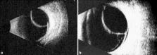
          The purpose of this paper is to report the case of a 62-year-old male diagnosed with a retinal macrocyst secondary to a long-standing retinal detachment in his right eye. At fundoscopy examination, an oval, elevated retinal lesion in the superior nasal quadrant was noted. Ultrasonography was performed, with a B-mode echography showing an oval, anechoic image and a standardized A-mode echography with a reflectivity spike higher than 98%, which was compatible with a retinal macrocyst. The patient refused surgical treatment for the retinal detachment and was followed for 14 months with stable visual acuity and no clinical or echographic changes.

          Related collections

          Most cited references5

          • Record: found
          • Abstract: found
          • Article: found
          Is Open Access

          Hemorrhagic intraretinal macrocyst: Differential diagnoses and report of an unusual case

          Retinal ‘cysts’ may be single or multiple, ranging from two-to-ten disc diameters in size, and occur in eyes with longstanding retinal detachment. The authors describe a retinal macrocyst larger than ten disc diameters, with a blood-filled cavity, and its ultrasound findings. Improved retinal nourishment following retinal reattachment gradually reverses the process responsible for cystic degeneration, with the eventual collapse of the cyst (within days or weeks). Surprisingly, this giant cyst did not collapse for almost three years despite retinal reattachment. The internal mobile echogenic contents were suggestive of blood. The possible reason of blood in the cyst could be rupture of the retinal blood vessels in the cyst cavity. This could be a recurrent phenomenon, which did not allow the cyst to collapse. The Hemorrhagic Intraretinal Macrocyst needs to be differentiated from mimicking clinical conditions, namely, retinoschisis, choroidal melanoma, subretinal abscess, choroidal hemangioma, and the like. It could take up to a few years to collapse spontaneously, following successful retinal reattachment.
            Bookmark
            • Record: found
            • Abstract: found
            • Article: not found

            Internal Drainage of a Retinal Macrocyst With an Nd:YAG Laser to Aid Primary Retinal Reattachment

            (2009)
            Retinal macrocysts are an occasional finding in patients with long-standing retinal detachments. The cysts usually do not require any specific treatment during procedures to repair retinal detachment and resolve spontaneously after retinal reattachment. However, if the cyst is adjacent to a retinal break and prevents its closure, the retinal reattachment procedure can fail. We describe a technique using the Nd:YAG laser to perforate the retinal cyst postoperatively, which allows for subsequent break closure and retinal reattachment.
              Bookmark
              • Record: found
              • Abstract: not found
              • Article: not found

              SURGICAL TREATMENT OF RETINAL CYSTS.

              K Pischel (1963)
                Bookmark

                Author and article information

                Journal
                Case Rep Ophthalmol
                Case Rep Ophthalmol
                COP
                Case Reports in Ophthalmology
                S. Karger AG (Allschwilerstrasse 10, P.O. Box · Postfach · Case postale, CH–4009, Basel, Switzerland · Schweiz · Suisse, Phone: +41 61 306 11 11, Fax: +41 61 306 12 34, karger@karger.ch )
                1663-2699
                May-Aug 2014
                5 June 2014
                5 June 2014
                : 5
                : 2
                : 168-171
                Affiliations
                Instituto de Oftalmologia ‘Conde de Valenciana’, Mexico City, Mexico
                Author notes
                *Juan Carlos Serna-Ojeda, MD, Instituto de Oftalmologia ‘Conde de Valenciana’, Chimalpopoca 14, Mexico City 06800 (Mexico), E-Mail juanc.sernao@ 123456gmail.com
                Article
                cop-0005-0168
                10.1159/000363759
                4086044
                3acfe5b5-0ced-4faa-8be6-696ea3388ade
                Copyright © 2014 by S. Karger AG, Basel

                This is an Open Access article licensed under the terms of the Creative Commons Attribution-NonCommercial 3.0 Unported license (CC BY-NC) (www.karger.com/OA-license), applicable to the online version of the article only. Users may download, print and share this work on the Internet for noncommercial purposes only, provided the original work is properly cited, and a link to the original work on http://www.karger.com and the terms of this license are included in any shared versions.

                History
                Page count
                Figures: 3, References: 5, Pages: 4
                Categories
                Published online: June, 2014

                retinal macrocyst,retinal detachment,b-mode echography,a-mode echography

                Comments

                Comment on this article