11
views
0
recommends
+1 Recommend
0 collections
    0
    shares
      • Record: found
      • Abstract: found
      • Article: found
      Is Open Access

      Comparison of strain imaging techniques in CRT candidates: CMR tagging, CMR feature tracking and speckle tracking echocardiography

      research-article

      Read this article at

          There is no author summary for this article yet. Authors can add summaries to their articles on ScienceOpen to make them more accessible to a non-specialist audience.

          Abstract

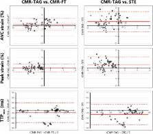
          Parameters using myocardial strain analysis may predict response to cardiac resynchronization therapy (CRT). As the agreement between currently available strain imaging modalities is unknown, three different modalities were compared. Twenty-seven CRT-candidates, prospectively included in the MARC study, underwent cardiac magnetic resonance (CMR) imaging and echocardiographic examination. Left ventricular (LV) circumferential strain was analysed with CMR tagging (CMR-TAG), CMR feature tracking (CMR-FT), and speckle tracking echocardiography (STE). Basic strain values and parameters of dyssynchrony and discoordination obtained with CMR-FT and STE were compared to CMR-TAG. Agreement of CMR-FT and CMR-TAG was overall fair, while agreement between STE and CMR-TAG was often poor. For both comparisons, agreement on discoordination parameters was highest, followed by dyssynchrony and basic strain parameters. For discoordination parameters, agreement on systolic stretch index was highest, with fair intra-class correlation coefficients (ICC) (CMR-FT: 0.58, STE: 0.55). ICC of septal systolic rebound stretch (SRS sept) was poor (CMR-FT: 0.41, STE: 0.30). Internal stretch factor of septal and lateral wall (ISF sep–lat) showed fair ICC values (CMR-FT: 0.53, STE: 0.46), while the ICC of the total LV (ISF LV) was fair for CMR-FT (0.55) and poor for STE (ICC: 0.32). The CURE index had a fair ICC for both comparisons (CMR-FT: 0.49, STE 0.41). Although comparison of STE to CMR-TAG was limited by methodological differences, agreement between CMR-FT and CMR-TAG was overall higher compared to STE and CMR-TAG. CMR-FT is a potential clinical alternative for CMR-TAG and STE, especially in the detection of discoordination in CRT-candidates.

          Electronic supplementary material

          The online version of this article (doi:10.1007/s10554-017-1253-5) contains supplementary material, which is available to authorized users.

          Related collections

          Most cited references28

          • Record: found
          • Abstract: found
          • Article: found
          Is Open Access

          Principles of cardiovascular magnetic resonance feature tracking and echocardiographic speckle tracking for informed clinical use

          Tissue tracking technology of routinely acquired cardiovascular magnetic resonance (CMR) cine acquisitions has increased the apparent ease and availability of non-invasive assessments of myocardial deformation in clinical research and practice. Its widespread availability thanks to the fact that this technology can in principle be applied on images that are part of every CMR or echocardiographic protocol. However, the two modalities are based on very different methods of image acquisition and reconstruction, each with their respective strengths and limitations. The image tracking methods applied are not necessarily directly comparable between the modalities, or with those based on dedicated CMR acquisitions for strain measurement such as tagging or displacement encoding. Here we describe the principles underlying the image tracking methods for CMR and echocardiography, and the translation of the resulting tracking estimates into parameters suited to describe myocardial mechanics. Technical limitations are presented with the objective of suggesting potential solutions that may allow informed and appropriate use in clinical applications.
            Bookmark
            • Record: found
            • Abstract: not found
            • Article: not found

            2012 EHRA/HRS expert consensus statement on cardiac resynchronization therapy in heart failure: implant and follow-up recommendations and management.

              Bookmark
              • Record: found
              • Abstract: found
              • Article: not found

              Circumferential and longitudinal strain in 3 myocardial layers in normal subjects and in patients with regional left ventricular dysfunction.

              The left ventricle is not homogenous and is composed of 3 myocardial layers. Until recently, magnetic resonance imaging has been the only noninvasive technique for detailed evaluation of the left ventricular (LV) wall. The aim of this study was to analyze strain in 3 myocardial layers using speckle-tracking echocardiography. Twenty normal subjects and 21 patients with LV dysfunction underwent echocardiography. Short-axis (for circumferential) and apical (for longitudinal strain) views were analyzed using modified speckle-tracking software enabling the analysis of strain in 3 myocardial layers. In normal subjects, longitudinal and circumferential strain was highest in the endocardium and lowest in the epicardium. Longitudinal endocardial and mid layer strain was highest in the apex and lowest in the base. Epicardial longitudinal strain was homogenous over the left ventricle. Circumferential 3-layer strain was highest in the apex and lowest in the base. In patients with LV dysfunction, strain was lower, with late diastolic or double peak. Three-layer analysis of circumferential and longitudinal strain using speckle-tracking imaging can be performed on a clinical basis and may become an important method for the assessment of real-time, quantitative global and regional LV function.
                Bookmark

                Author and article information

                Contributors
                +31-887550397 , w.m.vaneverdingen@umcutrecht.nl
                a.zweerink@vumc.nl
                Robin@Nijveldt.net
                o.a.e.salden@umcutrecht.nl
                m.meine@umcutrecht.nl
                a.h.maass@umcg.nl
                kevin.vernooy@mumc.nl
                f.j.delange@amc.uva.nl
                ac.vrossum@vumc.nl
                croisille@creatis.insa-lyon.fr
                patrick.clarysse@creatis.insa-lyon.fr
                b.geelhoed@umcg.nl
                m.rienstra@umcg.nl
                i.c.van.gelder@umcg.nl
                m.a.vos@umcutrecht.nl
                cp.allaart@vumc.nl
                m.j.m.cramer@umcutrecht.nl
                Journal
                Int J Cardiovasc Imaging
                Int J Cardiovasc Imaging
                The International Journal of Cardiovascular Imaging
                Springer Netherlands (Dordrecht )
                1569-5794
                1875-8312
                17 October 2017
                17 October 2017
                2018
                : 34
                : 3
                : 443-456
                Affiliations
                [1 ]ISNI 0000000090126352, GRID grid.7692.a, Department of Cardiology, , University Medical Centre Utrecht, ; Utrecht, The Netherlands
                [2 ]ISNI 0000 0004 0435 165X, GRID grid.16872.3a, Department of Cardiology, and Institute for Cardiovascular Research (ICaR-VU), , VU University Medical Centre, ; Amsterdam, The Netherlands
                [3 ]Department of Cardiology, Thoraxcenter, University of Groningen, University Medical Centre Groningen, Groningen, The Netherlands
                [4 ]ISNI 0000 0004 0480 1382, GRID grid.412966.e, Department of Cardiology, , Maastricht University Medical Centre, ; Maastricht, The Netherlands
                [5 ]ISNI 0000000404654431, GRID grid.5650.6, Department of Cardiology, , Academic Medical Centre, ; Amsterdam, The Netherlands
                [6 ]GRID grid.435013.0, Université Lyon, UJM-Saint-Etienne, INSA, CNRS UMR 5520, INSERM U1206, CREATIS, ; 42023 Saint-Etienne, France
                [7 ]ISNI 0000000120346234, GRID grid.5477.1, Department of Medical Physiology, , University of Utrecht, ; Utrecht, The Netherlands
                Article
                1253
                10.1007/s10554-017-1253-5
                5847211
                29043465
                88f1fd3e-33fc-4a79-b940-1ab58cc2f675
                © The Author(s) 2017

                Open AccessThis article is distributed under the terms of the Creative Commons Attribution 4.0 International License ( http://creativecommons.org/licenses/by/4.0/), which permits unrestricted use, distribution, and reproduction in any medium, provided you give appropriate credit to the original author(s) and the source, provide a link to the Creative Commons license, and indicate if changes were made.

                History
                : 14 August 2017
                : 29 September 2017
                Funding
                Funded by: FundRef http://dx.doi.org/10.13039/501100006020, Center for Translational Molecular Medicine;
                Award ID: 01C-203
                Categories
                Original Paper
                Custom metadata
                © Springer Science+Business Media B.V., part of Springer Nature 2018

                Cardiovascular Medicine
                strain,myocardial tagging,feature tracking,speckle tracking echocardiography,dyssynchrony,discoordination,cardiac resynchronization therapy

                Comments

                Comment on this article