32
views
0
recommends
+1 Recommend
0 collections
    0
    shares
      • Record: found
      • Abstract: found
      • Article: not found

      XIAP-mediated Caspase Inhibition in Hodgkin's Lymphoma–derived B Cells

      research-article

      Read this article at

      ScienceOpenPublisherPMC
          There is no author summary for this article yet. Authors can add summaries to their articles on ScienceOpen to make them more accessible to a non-specialist audience.

          Abstract

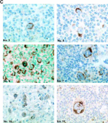
          The malignant Hodgkin and Reed-Sternberg cells of Hodgkin's lymphoma (HL) and HL-derived B cell lines were previously shown to be resistant to different apoptotic stimuli. We show here that cytochrome c fails to stimulate caspases-9 and -3 activation in cytosolic extracts of HL-derived B cells, which is due to high level expression of X-linked inhibitor of apoptosis (XIAP). Coimmunoprecipitation studies revealed that XIAP, apoptosis protease-activating factor–1, and caspase-3 are complexed in HL-derived B cell lysates. Even after stimulation with exogenous cytochrome c and dATP, XIAP impairs the proteolytic processing and activation of caspase-3. In cytosolic extracts, inhibition of XIAP by the second mitochondria-derived activator of caspases (Smac)/DIABLO, or immunodepletion of XIAP restores cytochrome c–triggered processing and activation of caspase-3. Smac or a Smac-derived agonistic peptide also sensitized intact HL-derived B cells for the apoptotic action of staurosporine. Finally, Hodgkin and Reed-Sternberg cells of primary tumor HL tissues also constitutively and abundantly express XIAP. The results of this paper suggest that high level XIAP expression is a hallmark of HL, which may play a crucial role in resistance to apoptosis.

          Related collections

          Most cited references20

          • Record: found
          • Abstract: found
          • Article: not found

          Cytochrome c and dATP-dependent formation of Apaf-1/caspase-9 complex initiates an apoptotic protease cascade.

          We report here the purification of the third protein factor, Apaf-3, that participates in caspase-3 activation in vitro. Apaf-3 was identified as a member of the caspase family, caspase-9. Caspase-9 and Apaf-1 bind to each other via their respective NH2-terminal CED-3 homologous domains in the presence of cytochrome c and dATP, an event that leads to caspase-9 activation. Activated caspase-9 in turn cleaves and activates caspase-3. Depletion of caspase-9 from S-100 extracts diminished caspase-3 activation. Mutation of the active site of caspase-9 attenuated the activation of caspase-3 and cellular apoptotic response in vivo, indicating that caspase-9 is the most upstream member of the apoptotic protease cascade that is triggered by cytochrome c and dATP.
            Bookmark
            • Record: found
            • Abstract: not found
            • Article: not found

            IAP family proteins--suppressors of apoptosis.

              Bookmark
              • Record: found
              • Abstract: found
              • Article: not found

              Two CD95 (APO-1/Fas) signaling pathways.

              We have identified two cell types, each using almost exclusively one of two different CD95 (APO-1/Fas) signaling pathways. In type I cells, caspase-8 was activated within seconds and caspase-3 within 30 min of receptor engagement, whereas in type II cells cleavage of both caspases was delayed for approximately 60 min. However, both type I and type II cells showed similar kinetics of CD95-mediated apoptosis and loss of mitochondrial transmembrane potential (DeltaPsim). Upon CD95 triggering, all mitochondrial apoptogenic activities were blocked by Bcl-2 or Bcl-xL overexpression in both cell types. However, in type II but not type I cells, overexpression of Bcl-2 or Bcl-xL blocked caspase-8 and caspase-3 activation as well as apoptosis. In type I cells, induction of apoptosis was accompanied by activation of large amounts of caspase-8 by the death-inducing signaling complex (DISC), whereas in type II cells DISC formation was strongly reduced and activation of caspase-8 and caspase-3 occurred following the loss of DeltaPsim. Overexpression of caspase-3 in the caspase-3-negative cell line MCF7-Fas, normally resistant to CD95-mediated apoptosis by overexpression of Bcl-xL, converted these cells into true type I cells in which apoptosis was no longer inhibited by Bcl-xL. In summary, in the presence of caspase-3 the amount of active caspase-8 generated at the DISC determines whether a mitochondria-independent apoptosis pathway is used (type I cells) or not (type II cells).
                Bookmark

                Author and article information

                Journal
                J Exp Med
                The Journal of Experimental Medicine
                The Rockefeller University Press
                0022-1007
                1540-9538
                21 July 2003
                : 198
                : 2
                : 341-347
                Affiliations
                [1 ]Institute for Medical Microbiology, Immunology, and Hygiene, University of Cologne, 50935 Köln, Germany
                [2 ]The Burnham Institute, Program for Apoptosis and Cell Death Research, La Jolla, CA 92037
                [3 ]Institute of Pathology, Aarhus University Hospital, Komunehospitalet, 8000 Aarhus C, Denmark
                Author notes

                Address correspondence to Martin Krönke, Institute for Medical Microbiology, Immunology and Hygiene, University of Cologne, Goldenfelsstrasse 19-21, 50935 Cologne, Germany. Phone: 49-221-478-3060; Fax: 49-221-478-3067; E-mail: martin.kroenke@ 123456medizin.uni-koeln.de

                Article
                20021279
                10.1084/jem.20021279
                2194071
                12874265
                16e29f23-9dad-416e-9e7a-a3582ca77010
                Copyright © 2003, The Rockefeller University Press
                History
                : 29 July 2002
                : 22 May 2003
                : 22 May 2003
                Categories
                Article

                Medicine
                mitochondria,cancer,tumor,smac/diablo,apoptosis
                Medicine
                mitochondria, cancer, tumor, smac/diablo, apoptosis

                Comments

                Comment on this article