- Record: found
- Abstract: found
- Article: found
Comment on “Clinical Comparisons of Two Free Light Chain Assays to Immunofixation Electrophoresis for Detecting Monoclonal Gammopathy”
letter
Read this article at
There is no author summary for this article yet. Authors can add summaries to their articles on ScienceOpen to make them more accessible to a non-specialist audience.
Abstract
The recent publication by Kim et al. [1] compares the performance of two serum free
light chain (sFLC) assays, the polyclonal antibody based Freelite and the monoclonal
antibody based N-Latex-FLC. We welcome the opportunity to comment on the design of
the study and interpretation of results.
Serum and urine electrophoresis can be used to identify monoclonal gammopathy (MG)
patients with gross intact monoclonal immunoglobulin or free light chain production.
However, electrophoresis assays are insensitive for the detection of patients with
AL amyloidosis and nonsecretory multiple myeloma (NSMM). The introduction of the Freelite
assay in 2001 [2] improved the sensitivity for detection of patients with monoclonal
free light chain production. This improved sensitivity has resulted in the inclusion
of Freelite in international guidelines [3–5] for screening, diagnosis, and monitoring
of monoclonal gammopathies. Recently, assays utilising monoclonal antibodies for the
measurement of serum free light chains have become available. The assays are calibrated
to Freelite, but so far there is a paucity of data comparing the clinical performance
of the assays.
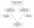
Kim et al. analysed samples from 63 MG (n = 100 samples) and 57 non-MG (n = 57 samples)
patients. Both kappa and lambda N-Latex-FLC are calibrated to the Freelite assays
[6] and therefore it is not surprising that there is concordance with results in a
normal population. However, we believe there are too few clinical samples from patients
with light chain multiple myeloma (LCMM) (10/63) and AL amyloidosis (2/63) for Kim
et al. to make a reliable clinical comparison.
There were 17 discordant results in this study (13 MG and 4 non-MG patients). It would
have been informative if the authors had presented the performance of the assays in
the different groups of monoclonal gammopathy patients, particularly in those with
LCMM and AL amyloidosis. Specifically in LCMM populations previous studies with the
monoclonal antibody based N-Latex-FLC test have failed to identify all patients [6–8].
By contrast in sixteen independent studies, including samples from 682 LCMM patients,
an abnormal κ/λ sFLC ratio using the Freelite assay identified 100% samples (Table
1) [9–25]. One study [26] reported a LCMM patient missed by Freelite; however the
sample was correctly identified when reanalysed using the same batch of reagent, indicating
a previous analytical error (personal communication). To truly understand the concordance
between the assays larger studies are required in clinically relevant populations
including patients with AL amyloidosis, LCMM, and NSMM. In addition, there has only
been a single study comparing the assays in patient with acute kidney injury [27].
4/57 non-MG patients had an abnormal κ/λ sFLC ratio using the Freelite assay but had
normal ratios using the N-Latex-FLC assay. These patients had disorders (chronic kidney
disease, chronic obstructive pulmonary disease, iron deficiency anaemia, and systemic
lupus erythematosus) that have previously been reported to cause a perturbation of
the κ/λ sFLC ratio due to poor renal function, inflammation, or immune stimulation
[28–30]. In patients with renal impairment FLC removal becomes increasingly dependent
on the reticuloendothelial system. Unlike renal clearance reticuloendothelial clearance
is not influenced by size of the light chains [31]; therefore the production rate
of kappa FLC (approximately 2x that of lambda) exerts an influence on the κ/λ FLC
ratio. Whilst there have previously been reports highlighting the difference in the
performance of the N-Latex-FLC assay in patients with impaired renal function, there
has been no physiological explanation for this performance [32].
The quantitative assessment of free light chains by Freelite is an important laboratory
test. An abnormal ratio can be used as part of an algorithm to risk stratifying monoclonal
gammopathy of undetermined significance patients. Furthermore, a ratio of >100 with
a monoclonal free light chain concentration >100 mg/L was recently included in the
diagnostic algorithm for patients with multiple myeloma, and an abnormal ratio is
useful in understanding the depth of response in patients during the course of their
disease [33–35]. Given the reliance upon the numerical values we believe there is
a strong requirement for better quantitative concordance between the assays, and clearly
the role of Freelite in diagnosis, stratification, and response cannot be transferred
to the N-Latex-FLC assay.
In summary, sample selection in this study limits interpretation but supports published
data showing that differences exist between the polyclonal and monoclonal FLC assays.
Related collections
Most cited references32
- Record: found
- Abstract: found
- Article: not found
Highly sensitive, automated immunoassay for immunoglobulin free light chains in serum and urine.
Richard Drew, Daniel G. Mead, A R Bradwell … (2001)
- Record: found
- Abstract: found
- Article: found
Serum free light chain measurement aids the diagnosis of myeloma in patients with severe renal failure
Colin A. Hutchison, Tim Plant, Mark Drayson … (2008)
- Record: found
- Abstract: found
- Article: not found
Importance of achieving stringent complete response after autologous stem-cell transplantation in multiple myeloma.
Prashant Kapoor, Shaji K Kumar, Angela Dispenzieri … (2013)