0
views
0
recommends
+1 Recommend
0 collections
    0
    shares
      • Record: found
      • Abstract: found
      • Article: found
      Is Open Access

      Pathophysiology of reversible cerebral vasoconstriction syndrome

      review-article

      Read this article at

          There is no author summary for this article yet. Authors can add summaries to their articles on ScienceOpen to make them more accessible to a non-specialist audience.

          Abstract

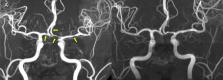
          Reversible cerebral vasoconstriction syndrome (RCVS) is a complex neurovascular disorder being recognized during the past two decades. It is characterized by multiple abrupt severe headaches and widespread cerebral vasoconstrictions, with potential complications such as ischemic stroke, convexity subarachnoid hemorrhage, intracerebral hemorrhage and posterior reversible encephalopathy syndrome. The clinical features, imaging findings, and dynamic disease course have been delineated. However, the pathophysiology of RCVS remains elusive. Recent studies have had substantial progress in elucidating its pathogenesis. It is now believed that dysfunction of cerebral vascular tone and impairment of blood–brain barrier may play key roles in the pathophysiology of RCVS, which explains some of the clinical and radiological manifestations of RCVS. Some other potentially important elements include genetic predisposition, sympathetic overactivity, endothelial dysfunction, and oxidative stress, although the detailed molecular mechanisms are yet to be identified. In this review, we will summarize what have been revealed in the literature and elaborate how these factors could contribute to the pathophysiology of RCVS.

          Related collections

          Most cited references121

          • Record: found
          • Abstract: found
          • Article: not found

          Structure and function of the blood-brain barrier.

          Neural signalling within the central nervous system (CNS) requires a highly controlled microenvironment. Cells at three key interfaces form barriers between the blood and the CNS: the blood-brain barrier (BBB), blood-CSF barrier and the arachnoid barrier. The BBB at the level of brain microvessel endothelium is the major site of blood-CNS exchange. The structure and function of the BBB is summarised, the physical barrier formed by the endothelial tight junctions, and the transport barrier resulting from membrane transporters and vesicular mechanisms. The roles of associated cells are outlined, especially the endfeet of astrocytic glial cells, and pericytes and microglia. The embryonic development of the BBB, and changes in pathology are described. The BBB is subject to short and long-term regulation, which may be disturbed in pathology. Any programme for drug discovery or delivery, to target or avoid the CNS, needs to consider the special features of the BBB.
            Bookmark
            • Record: found
            • Abstract: found
            • Article: not found

            Circulating angiogenic factors and the risk of preeclampsia.

            The cause of preeclampsia remains unclear. Limited data suggest that excess circulating soluble fms-like tyrosine kinase 1 (sFlt-1), which binds placental growth factor (PlGF) and vascular endothelial growth factor (VEGF), may have a pathogenic role. We performed a nested case-control study within the Calcium for Preeclampsia Prevention trial, which involved healthy nulliparous women. Each woman with preeclampsia was matched to one normotensive control. A total of 120 pairs of women were randomly chosen. Serum concentrations of angiogenic factors (total sFlt-1, free PlGF, and free VEGF) were measured throughout pregnancy; there were a total of 655 serum specimens. The data were analyzed cross-sectionally within intervals of gestational age and according to the time before the onset of preeclampsia. During the last two months of pregnancy in the normotensive controls, the level of sFlt-1 increased and the level of PlGF decreased. These changes occurred earlier and were more pronounced in the women in whom preeclampsia later developed. The sFlt-1 level increased beginning approximately five weeks before the onset of preeclampsia. At the onset of clinical disease, the mean serum level in the women with preeclampsia was 4382 pg per milliliter, as compared with 1643 pg per milliliter in controls with fetuses of similar gestational age (P<0.001). The PlGF levels were significantly lower in the women who later had preeclampsia than in the controls beginning at 13 to 16 weeks of gestation (mean, 90 pg per milliliter vs. 142 pg per milliliter, P=0.01), with the greatest difference occurring during the weeks before the onset of preeclampsia, coincident with the increase in the sFlt-1 level. Alterations in the levels of sFlt-1 and free PlGF were greater in women with an earlier onset of preeclampsia and in women in whom preeclampsia was associated with a small-for-gestational-age infant. Increased levels of sFlt-1 and reduced levels of PlGF predict the subsequent development of preeclampsia. Copyright 2004 Massachusetts Medical Society
              Bookmark
              • Record: found
              • Abstract: found
              • Article: not found

              Endothelial progenitor cells: characterization and role in vascular biology.

              Infusion of different hematopoietic stem cell populations and ex vivo expanded endothelial progenitor cells augments neovascularization of tissue after ischemia and contributes to reendothelialization after endothelial injury, thereby, providing a novel therapeutic option. However, controversy exists with respect to the identification and the origin of endothelial progenitor cells. Overall, there is consensus that endothelial progenitor cells can derive from the bone marrow and that CD133/VEGFR2 cells represent a population with endothelial progenitor capacity. However, increasing evidence suggests that there are additional bone marrow-derived cell populations (eg, myeloid cells, "side population" cells, and mesenchymal cells) and non-bone marrow-derived cells, which also can give rise to endothelial cells. The characterization of the different progenitor cell populations and their functional properties are discussed. Mobilization and endothelial progenitor cell-mediated neovascularization is critically regulated. Stimulatory (eg, statins and exercise) or inhibitory factors (risk factors for coronary artery disease) modulate progenitor cell levels and, thereby, affect the vascular repair capacity. Moreover, recruitment and incorporation of endothelial progenitor cells requires a coordinated sequence of multistep adhesive and signaling events including adhesion and migration (eg, by integrins), chemoattraction (eg, by SDF-1/CXCR4), and finally the differentiation to endothelial cells. This review summarizes the mechanisms regulating endothelial progenitor cell-mediated neovascularization and reendothelialization.
                Bookmark

                Author and article information

                Contributors
                chensp1977@gmail.com
                sjwang@vghtpe.gov.tw
                Journal
                J Biomed Sci
                J Biomed Sci
                Journal of Biomedical Science
                BioMed Central (London )
                1021-7770
                1423-0127
                21 September 2022
                21 September 2022
                2022
                : 29
                : 72
                Affiliations
                [1 ]GRID grid.278247.c, ISNI 0000 0004 0604 5314, Department of Neurology, , Neurological Institute, Taipei Veterans General Hospital, ; Taipei, 11217 Taiwan
                [2 ]GRID grid.260539.b, ISNI 0000 0001 2059 7017, Institute of Clinical Medicine, , National Yang Ming Chiao Tung University, ; Taipei, Taiwan
                [3 ]GRID grid.278247.c, ISNI 0000 0004 0604 5314, Division of Translational Research, Department of Medical Research, , Taipei Veterans General Hospital, ; Taipei, Taiwan
                [4 ]GRID grid.260539.b, ISNI 0000 0001 2059 7017, Brain Research Center & School of Medicine, , National Yang Ming Chiao Tung University, ; Taipei, Taiwan
                Author information
                http://orcid.org/0000-0003-3492-9902
                Article
                857
                10.1186/s12929-022-00857-4
                9489486
                36127720
                7bcd3524-ae74-4fff-a751-2a6c15653ee5
                © The Author(s) 2022

                Open AccessThis article is licensed under a Creative Commons Attribution 4.0 International License, which permits use, sharing, adaptation, distribution and reproduction in any medium or format, as long as you give appropriate credit to the original author(s) and the source, provide a link to the Creative Commons licence, and indicate if changes were made. The images or other third party material in this article are included in the article's Creative Commons licence, unless indicated otherwise in a credit line to the material. If material is not included in the article's Creative Commons licence and your intended use is not permitted by statutory regulation or exceeds the permitted use, you will need to obtain permission directly from the copyright holder. To view a copy of this licence, visit http://creativecommons.org/licenses/by/4.0/. The Creative Commons Public Domain Dedication waiver ( http://creativecommons.org/publicdomain/zero/1.0/) applies to the data made available in this article, unless otherwise stated in a credit line to the data.

                History
                : 28 July 2022
                : 14 September 2022
                Funding
                Funded by: FundRef http://dx.doi.org/10.13039/100010002, Ministry of Education;
                Award ID: The Featured Areas Research Center Program within the framework of the Higher Education Sprout Project by the Ministry of Education (MOE) in Taiwan
                Award Recipient :
                Funded by: Ministry of Science and Technology (TW)
                Award ID: MOST-107-2314-B-010-021
                Award ID: 108-2314-B-010-022-MY3
                Award ID: 110-2326-B-A49A-501-MY3
                Award Recipient :
                Funded by: FundRef http://dx.doi.org/10.13039/501100004663, Ministry of Science and Technology, Taiwan;
                Award ID: MOST 108-2321-B-010-014-MY2
                Award ID: 108-2321-B-010-001-
                Award ID: 108-2314-B-010-023-MY3
                Award ID: 110-2321-B-010-005-
                Award ID: 111-2321-B-A49-004-
                Award Recipient :
                Funded by: FundRef http://dx.doi.org/10.13039/501100011912, Taipei Veterans General Hospital;
                Award ID: V111C-158
                Award ID: V109D52-001-MY3-3
                Award ID: VGHUST110-G1-3-1
                Award ID: VGH-106-D9-001-MY2-2
                Award Recipient :
                Categories
                Review
                Custom metadata
                © The Author(s) 2022

                Molecular medicine
                reversible cerebral vasoconstriction syndrome,thunderclap headache,blood–brain barrier,neurovascular unit

                Comments

                Comment on this article