6
views
0
recommends
+1 Recommend
0 collections
    0
    shares
      • Record: found
      • Abstract: found
      • Article: found
      Is Open Access

      High Expression of Cancer-IgG Is Associated With Poor Prognosis and Radioresistance via PI3K/AKT/DNA-PKcs Pathway Regulation in Lung Adenocarcinoma

      research-article

      Read this article at

      Bookmark
          There is no author summary for this article yet. Authors can add summaries to their articles on ScienceOpen to make them more accessible to a non-specialist audience.

          Abstract

          Background

          Lung adenocarcinoma (LUAD) is the dominant type of lung neoplasms, and radiotherapy is its mainstay treatment, yet poor prognosis caused by radioresistance remains problematic. Cancer-derived immunoglobulin G (cancer-IgG) has been detected in multiple cancers and plays important roles in carcinogenesis. This study aimed to demonstrate that cancer-IgG is associated with poor prognosis of LUAD and to identify its role in radioresistance.

          Methods

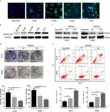
          Cancer-IgG expression was detected by immunohistochemistry from 56 patients with stage III LUAD and by western blot and immunofluorescence in LUAD cell lines and in a human bronchial epithelial cell line. The effects of cancer-IgG silencing on the proliferation and apoptosis of PC9 and H292 cells were evaluated by plate cloning and apoptosis assay; the effects of cancer-IgG silencing on DNA damage repair ability and radiosensitivity were evaluated by colony-forming assay, γH2AX immunofluorescence, and neutral comet assay. Finally, we used the protein phosphorylation microarray and western blot to explore mechanisms involving cancer-IgG that increased radioresistance.

          Results

          Cancer-IgG is widely expressed in stage III LUAD, and the overall survival and disease-free survival of patients with positive expression are notably lower than those of patients with negative expression, indicating the associations between cancer-IgG and poor prognosis as well as radioresistance. The expression of cancer-IgG in the four LUAD cell lines was located mainly on the cell membrane and cytoplasm and not in the normal lung epithelial cell. Knockdown of cancer-IgG in PC9 and H292 cells resulted in increased apoptosis and negatively affected cancer cell proliferation. After irradiation, silencing of cancer-IgG showed a decrease in colonies as well as increases in the Olive tail moment and γH2AX foci in nucleus, indicating that the knockdown of cancer-IgG resulted in a decrease in the damage repair ability of DNA double-strand breaks in LUAD cells and an enhanced radiosensitivity. The expression of p-AKT, p-GSK3β, and p-DNA-PKcs decreased in the knockdown group after radiotherapy, suggesting that cancer-IgG could affect radiotherapy resistance by mediating double-strand breaks damage repair in LUAD cells through the PI3K/AKT/DNA-PKcs pathway.

          Conclusions

          This study revealed that cancer-IgG regulates PI3K/AKT/DNA-PKcs signaling pathways to affect radioresistance of LUAD and associated with poor prognosis.

          Related collections

          Most cited references37

          • Record: found
          • Abstract: found
          • Article: not found

          Cancer statistics, 2020

          Each year, the American Cancer Society estimates the numbers of new cancer cases and deaths that will occur in the United States and compiles the most recent data on population-based cancer occurrence. Incidence data (through 2016) were collected by the Surveillance, Epidemiology, and End Results Program; the National Program of Cancer Registries; and the North American Association of Central Cancer Registries. Mortality data (through 2017) were collected by the National Center for Health Statistics. In 2020, 1,806,590 new cancer cases and 606,520 cancer deaths are projected to occur in the United States. The cancer death rate rose until 1991, then fell continuously through 2017, resulting in an overall decline of 29% that translates into an estimated 2.9 million fewer cancer deaths than would have occurred if peak rates had persisted. This progress is driven by long-term declines in death rates for the 4 leading cancers (lung, colorectal, breast, prostate); however, over the past decade (2008-2017), reductions slowed for female breast and colorectal cancers, and halted for prostate cancer. In contrast, declines accelerated for lung cancer, from 3% annually during 2008 through 2013 to 5% during 2013 through 2017 in men and from 2% to almost 4% in women, spurring the largest ever single-year drop in overall cancer mortality of 2.2% from 2016 to 2017. Yet lung cancer still caused more deaths in 2017 than breast, prostate, colorectal, and brain cancers combined. Recent mortality declines were also dramatic for melanoma of the skin in the wake of US Food and Drug Administration approval of new therapies for metastatic disease, escalating to 7% annually during 2013 through 2017 from 1% during 2006 through 2010 in men and women aged 50 to 64 years and from 2% to 3% in those aged 20 to 49 years; annual declines of 5% to 6% in individuals aged 65 years and older are particularly striking because rates in this age group were increasing prior to 2013. It is also notable that long-term rapid increases in liver cancer mortality have attenuated in women and stabilized in men. In summary, slowing momentum for some cancers amenable to early detection is juxtaposed with notable gains for other common cancers.
            Bookmark
            • Record: found
            • Abstract: found
            • Article: found
            Is Open Access

            Estimates and 25-year trends of the global burden of disease attributable to ambient air pollution: an analysis of data from the Global Burden of Diseases Study 2015

            Summary Background Exposure to ambient air pollution increases morbidity and mortality, and is a leading contributor to global disease burden. We explored spatial and temporal trends in mortality and burden of disease attributable to ambient air pollution from 1990 to 2015 at global, regional, and country levels. Methods We estimated global population-weighted mean concentrations of particle mass with aerodynamic diameter less than 2·5 μm (PM2·5) and ozone at an approximate 11 km × 11 km resolution with satellite-based estimates, chemical transport models, and ground-level measurements. Using integrated exposure–response functions for each cause of death, we estimated the relative risk of mortality from ischaemic heart disease, cerebrovascular disease, chronic obstructive pulmonary disease, lung cancer, and lower respiratory infections from epidemiological studies using non-linear exposure–response functions spanning the global range of exposure. Findings Ambient PM2·5 was the fifth-ranking mortality risk factor in 2015. Exposure to PM2·5 caused 4·2 million (95% uncertainty interval [UI] 3·7 million to 4·8 million) deaths and 103·1 million (90·8 million 115·1 million) disability-adjusted life-years (DALYs) in 2015, representing 7·6% of total global deaths and 4·2% of global DALYs, 59% of these in east and south Asia. Deaths attributable to ambient PM2·5 increased from 3·5 million (95% UI 3·0 million to 4·0 million) in 1990 to 4·2 million (3·7 million to 4·8 million) in 2015. Exposure to ozone caused an additional 254 000 (95% UI 97 000–422 000) deaths and a loss of 4·1 million (1·6 million to 6·8 million) DALYs from chronic obstructive pulmonary disease in 2015. Interpretation Ambient air pollution contributed substantially to the global burden of disease in 2015, which increased over the past 25 years, due to population ageing, changes in non-communicable disease rates, and increasing air pollution in low-income and middle-income countries. Modest reductions in burden will occur in the most polluted countries unless PM2·5 values are decreased substantially, but there is potential for substantial health benefits from exposure reduction. Funding Bill & Melinda Gates Foundation and Health Effects Institute.
              Bookmark
              • Record: found
              • Abstract: found
              • Article: not found

              Durvalumab after Chemoradiotherapy in Stage III Non-Small-Cell Lung Cancer.

              Background Most patients with locally advanced, unresectable, non-small-cell lung cancer (NSCLC) have disease progression despite definitive chemoradiotherapy (chemotherapy plus concurrent radiation therapy). This phase 3 study compared the anti-programmed death ligand 1 antibody durvalumab as consolidation therapy with placebo in patients with stage III NSCLC who did not have disease progression after two or more cycles of platinum-based chemoradiotherapy. Methods We randomly assigned patients, in a 2:1 ratio, to receive durvalumab (at a dose of 10 mg per kilogram of body weight intravenously) or placebo every 2 weeks for up to 12 months. The study drug was administered 1 to 42 days after the patients had received chemoradiotherapy. The coprimary end points were progression-free survival (as assessed by means of blinded independent central review) and overall survival (unplanned for the interim analysis). Secondary end points included 12-month and 18-month progression-free survival rates, the objective response rate, the duration of response, the time to death or distant metastasis, and safety. Results Of 713 patients who underwent randomization, 709 received consolidation therapy (473 received durvalumab and 236 received placebo). The median progression-free survival from randomization was 16.8 months (95% confidence interval [CI], 13.0 to 18.1) with durvalumab versus 5.6 months (95% CI, 4.6 to 7.8) with placebo (stratified hazard ratio for disease progression or death, 0.52; 95% CI, 0.42 to 0.65; P<0.001); the 12-month progression-free survival rate was 55.9% versus 35.3%, and the 18-month progression-free survival rate was 44.2% versus 27.0%. The response rate was higher with durvalumab than with placebo (28.4% vs. 16.0%; P<0.001), and the median duration of response was longer (72.8% vs. 46.8% of the patients had an ongoing response at 18 months). The median time to death or distant metastasis was longer with durvalumab than with placebo (23.2 months vs. 14.6 months; P<0.001). Grade 3 or 4 adverse events occurred in 29.9% of the patients who received durvalumab and 26.1% of those who received placebo; the most common adverse event of grade 3 or 4 was pneumonia (4.4% and 3.8%, respectively). A total of 15.4% of patients in the durvalumab group and 9.8% of those in the placebo group discontinued the study drug because of adverse events. Conclusions Progression-free survival was significantly longer with durvalumab than with placebo. The secondary end points also favored durvalumab, and safety was similar between the groups. (Funded by AstraZeneca; PACIFIC ClinicalTrials.gov number, NCT02125461 .).
                Bookmark

                Author and article information

                Contributors
                Journal
                Front Oncol
                Front Oncol
                Front. Oncol.
                Frontiers in Oncology
                Frontiers Media S.A.
                2234-943X
                02 June 2021
                2021
                : 11
                : 675397
                Affiliations
                [1] 1 Department of Radiation Oncology, Peking University China-Japan Friendship School of Clinical Medicine , Beijing, China
                [2] 2 Department of Radiotherapy, Second Hospital of Hebei Medical University , Shijiazhuang, China
                [3] 3 Key Laboratory of Carcinogenesis and Translational Research (Ministry of Education), Department of Radiation Oncology, Peking University Cancer Hospital and Institute , Beijing, China
                [4] 4 Department of Radiation Oncology, China-Japan Friendship Institute of Clinical Medicine , Beijing, China
                [5] 5 Department of Radiation Oncology, Center of Respiratory Medicine, China-Japan Friendship Hospital , Beijing, China
                [6] 6 Institute of Respiratory Medicine, Chinese Academy of Medical Sciences , Beijing, China
                [7] 7 Department of Radiation Oncology, National Clinical Research Center for Respiratory Diseases , Beijing, China
                [8] 8 Department of Radiation Oncology, World Health Organization (WHO) Collaborating Centre for Tobacco Cessation and Respiratory Diseases Prevention , Beijing, China
                [9] 9 Department of Pathology, China-Japan Friendship Hospital , Beijing, China
                [10] 10 Department of Immunology, School of Basic Medical Sciences, Peking University , Beijing, China
                [11] 11 Peking University Center for Human Disease Genomics , Beijing, China
                Author notes

                Edited by: Shubhankar Suman, Georgetown University, United States

                Reviewed by: Kamendra Kumar, Georgetown University, United States; Raghubendra Singh Dagur, University of Nebraska Medical Center, United States

                *Correspondence: Xiaoyan Qiu, qiuxy@ 123456bjmu.edu.cn ; Guangying Zhu, zryyfa@ 123456163.com

                †These authors have contributed equally to this work and share first authorship

                This article was submitted to Radiation Oncology, a section of the journal Frontiers in Oncology

                Article
                10.3389/fonc.2021.675397
                8208032
                34150640
                004c489b-6bed-4906-af21-ca921914ca0b
                Copyright © 2021 Yang, Wang, You, Gu, Xu, Xu, Wang, Zhao, Qiu and Zhu

                This is an open-access article distributed under the terms of the Creative Commons Attribution License (CC BY). The use, distribution or reproduction in other forums is permitted, provided the original author(s) and the copyright owner(s) are credited and that the original publication in this journal is cited, in accordance with accepted academic practice. No use, distribution or reproduction is permitted which does not comply with these terms.

                History
                : 03 March 2021
                : 19 April 2021
                Page count
                Figures: 5, Tables: 1, Equations: 0, References: 37, Pages: 10, Words: 5038
                Categories
                Oncology
                Original Research

                Oncology & Radiotherapy
                cancer-igg,lung adenocarcinoma,radioresistance,pi3k/akt/dna-pkcs pathway,rp215

                Comments

                Comment on this article