Inviting an author to review:
Find an author and click ‘Invite to review selected article’ near their name.
Search for authorsSearch for similar articles
1
views
0
recommends
+1 Recommend
0 collections
    0
    shares
      • Record: found
      • Abstract: not found
      • Article: not found

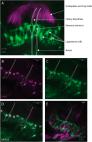
      Peripheral sensory cells in the cephalic sensory organs of Lymnaea stagnalis

      ,
      The Journal of Comparative Neurology
      Wiley

      Read this article at

      ScienceOpenPublisherPubMed
      Bookmark
          There is no author summary for this article yet. Authors can add summaries to their articles on ScienceOpen to make them more accessible to a non-specialist audience.

          Related collections

          Most cited references70

          • Record: found
          • Abstract: found
          • Article: not found

          User-friendly semiautomated assembly of accurate image mosaics in microscopy.

          We present a semiautomated software solution to the problem of extending the lateral field of view of a classical microscope. The initial requirements are a set of overlapping images, along with their user-provided coarse mosaic. Our solution then refines this initial mosaic in a fully automatic fashion. We rely on a highly accurate registration engine to perform the pairwise registration of the individual images, and on an efficient strategy to minimize the amount of computations while maintaining the highest possible global quality. We describe these ingredients, which we make available as a free multiplatform user-friendly software package. We also highlight why and how the specific aspects of the present microscopy application differ from those encountered while creating more common mosaics such as panoramas. Finally, we present experimental results that illustrate and validate our method on a real biological sample. We conclude by showing that we are able to reach subpixel accuracy.
            Bookmark
            • Record: found
            • Abstract: found
            • Article: not found

            Nitric oxide synthase and neuronal NADPH diaphorase are identical in brain and peripheral tissues.

            NADPH diaphorase staining neurons, uniquely resistant to toxic insults and neurodegenerative disorders, have been colocalized with neurons in the brain and peripheral tissue containing nitric oxide synthase (EC 1.14.23.-), which generates nitric oxide (NO), a recently identified neuronal messenger molecule. In the corpus striatum and cerebral cortex, NO synthase immunoreactivity and NADPH diaphorase staining are colocalized in medium to large aspiny neurons. These same neurons colocalize with somatostatin and neuropeptide Y immunoreactivity. NO synthase immunoreactivity and NADPH diaphorase staining are colocalized in the pedunculopontine nucleus with choline acetyltransferase-containing cells and are also colocalized in amacrine cells of the inner nuclear layer and ganglion cells of the retina, myenteric plexus neurons of the intestine, and ganglion cells of the adrenal medulla. Transfection of human kidney cells with NO synthase cDNA elicits NADPH diaphorase staining. The ratio of NO synthase to NADPH diaphorase staining in the transfected cells is the same as in neurons, indicating that NO synthase fully accounts for observed NADPH staining. The identity of neuronal NO synthase and NADPH diaphorase suggests a role for NO in modulating neurotoxicity.
              Bookmark
              • Record: found
              • Abstract: found
              • Article: not found

              Structure and innervation of the cochlea.

              The role of the cochlea is to transduce complex sound waves into electrical neural activity in the auditory nerve. Hair cells of the organ of Corti are the sensory cells of hearing. The inner hair cells perform the transduction and initiate the depolarization of the spiral ganglion neurons. The outer hair cells are accessory sensory cells that enhance the sensitivity and selectivity of the cochlea. Neural feedback loops that bring efferent signals to the outer hair cells assist in sharpening and amplifying the signals. The stria vascularis generates the endocochlear potential and maintains the ionic composition of the endolymph, the fluid in which the apical surface of the hair cells is bathed. The mechanical characteristics of the basilar membrane and its related structures further enhance the frequency selectivity of the auditory transduction mechanism. The tectorial membrane is an extracellular matrix, which provides mass loading on top of the organ of Corti, facilitating deflection of the stereocilia. This review deals with the structure of the normal mature mammalian cochlea and includes recent data on the molecular organization of the main cell types within the cochlea.
                Bookmark

                Author and article information

                Journal
                The Journal of Comparative Neurology
                J. Comp. Neurol.
                Wiley
                00219967
                July 01 2011
                July 01 2011
                May 23 2011
                : 519
                : 10
                : 1894-1913
                Article
                10.1002/cne.22607
                21452209
                00f7450c-8315-4fbf-8d36-8faba0e8c1ee
                © 2011

                http://doi.wiley.com/10.1002/tdm_license_1

                History

                Comments

                Comment on this article