0
views
0
recommends
+1 Recommend
0 collections
    0
    shares
      • Record: found
      • Abstract: found
      • Article: found
      Is Open Access

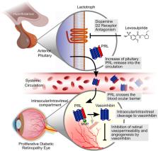
      Levosulpiride Increases the Levels of Prolactin and Antiangiogenic Vasoinhibin in the Vitreous of Patients with Proliferative Diabetic Retinopathy

      research-article

      Read this article at

      Bookmark
          There is no author summary for this article yet. Authors can add summaries to their articles on ScienceOpen to make them more accessible to a non-specialist audience.

          Abstract

          Purpose

          High circulating levels of the hormone prolactin (PRL) protect against experimental diabetic retinopathy (DR) due to the retinal accumulation of vasoinhibin, a PRL fragment that inhibits blood vessel permeability and growth. A phase 2 clinical trial is investigating a new therapy for DR based on elevating serum PRL levels with levosulpiride, a prokinetic dopamine D2 receptor blocker. Here, we tested whether levosulpiride-induced hyperprolactinemia elevates PRL and vasoinhibin in the vitreous of volunteer patients with proliferative DR (PDR) undergoing elective pars plana vitrectomy.

          Methods

          Patients were randomized to receive placebo (lactose pill, orally TID; n = 19) or levosulpiride (25 mg orally TID; n = 18) for the 7 days before vitrectomy. Vitreous samples from untreated non-diabetic ( n = 10) and PDR ( n = 17) patients were also studied.

          Results

          Levosulpiride elevated the systemic (101 ± 13 [SEM] vs. 9.2 ± 1.3 ng/mL, P < 0.0001) and vitreous (3.2 ± 0.4 vs. 1.5 ± 0.2 ng/mL, P < 0.0001) levels of PRL, and both levels were directly correlated ( r = 0.58, P < 0.0002). The vitreous from non-diabetic patients or from PDR patients treated with levosulpiride, but not from placebo-treated PDR patients, inhibited the basic fibroblast growth factor (bFGF)- and vascular endothelial growth factor (VEGF)-induced proliferation of endothelial cells in culture. Vasoinhibin-neutralizing antibodies reduced the vitreous antiangiogenic effect. Matrix metalloproteases (MMPs) in the vitreous cleaved PRL to vasoinhibin, and their activity was higher in non-diabetic than in PDR patients.

          Conclusions

          Levosulpiride increases the levels of PRL in the vitreous of PDR patients and promotes its MMP-mediated conversion to vasoinhibin, which can inhibit angiogenesis in DR.

          Translational Relevance

          These findings support the potential therapeutic benefit of levosulpiride against vision loss in diabetes. 

          Related collections

          Most cited references58

          • Record: found
          • Abstract: found
          • Article: not found

          A protocol for isolation and culture of human umbilical vein endothelial cells.

          We describe a protocol for easy isolation and culture of human umbilical vein endothelial cells (HUVECs) to supply every researcher with a method that can be applied in cell biology laboratories with minimum equipment. Endothelial cells (ECs) are isolated from umbilical vein vascular wall by a collagenase treatment, then seeded on fibronectin-coated plates and cultured in a medium with Earles' salts and fetal calf serum (FCS), but without growth factor supplementation, for 7 days in a 37 degrees C-5% CO2 incubator. Cell confluency can be monitored by phase-contrast microscopy; ECs can be characterized using cell surface or intracellular markers and checked for contamination. Various protocols can be applied to HUVECs, from simple harvesting to a particular solubilization of proteins for proteomic analysis.
            Bookmark
            • Record: found
            • Abstract: found
            • Article: not found

            The retina in Parkinson's disease.

            As a more complete picture of the clinical phenotype of Parkinson's disease emerges, non-motor symptoms have become increasingly studied. Prominent among these non-motor phenomena are mood disturbance, cognitive decline and dementia, sleep disorders, hyposmia and autonomic failure. In addition, visual symptoms are common, ranging from complaints of dry eyes and reading difficulties, through to perceptual disturbances (feelings of presence and passage) and complex visual hallucinations. Such visual symptoms are a considerable cause of morbidity in Parkinson's disease and, with respect to visual hallucinations, are an important predictor of cognitive decline as well as institutional care and mortality. Evidence exists of visual dysfunction at several levels of the visual pathway in Parkinson's disease. This includes psychophysical, electrophysiological and morphological evidence of disruption of retinal structure and function, in addition to disorders of 'higher' (cortical) visual processing. In this review, we will draw together work from animal and human studies in an attempt to provide an insight into how Parkinson's disease affects the retina and how these changes might contribute to the visual symptoms experienced by patients.
              Bookmark
              • Record: found
              • Abstract: found
              • Article: not found

              Ocular neovascularization: Implication of endogenous angiogenic inhibitors and potential therapy.

              Ocular neovascularization (NV) is the primary cause of blindness in a wide range of ocular diseases, such as diabetic retinopathy (DR) and age-related macular degeneration (AMD). The exact mechanism underlying the pathogenesis of ocular NV is not yet well understood, and as a consequence, there is no satisfactory therapy for ocular NV. In the last 10 years, a number of studies provided increasing evidence demonstrating that the imbalance between angiogenic stimulating factors and angiogenic inhibitors is a major contributor to the angiogenesis induced by various insults, such as hypoxia or ischemia, inflammation and tumor. The angiogenic inhibitors alone or in combination with other existing therapies are, therefore, believed to be promising in the treatment of ocular NV in the near future. This article reviews recent progress in studies on the mechanisms and treatment of ocular NV, focusing on the implication and therapeutic potential of endogenous angiogenic inhibitors in ocular NV.
                Bookmark

                Author and article information

                Journal
                Transl Vis Sci Technol
                Transl Vis Sci Technol
                tvst
                TVST
                Translational Vision Science & Technology
                The Association for Research in Vision and Ophthalmology
                2164-2591
                17 August 2020
                August 2020
                : 9
                : 9
                : 27
                Affiliations
                [1 ]Facultad de Ciencias Naturales, Universidad Autónoma de Querétaro, Querétaro, México
                [2 ]Instituto de Neurobiología, Universidad Nacional Autónoma de México, Campus Juriquilla, Querétaro, México
                [3 ]Instituto de la Retina del Bajío, Querétaro, México
                [4 ]Instituto Mexicano de Oftalmología, Querétaro, México
                [5 ]Institute for Clinical Chemistry, Laboratory Medicine and Transfusion Medicine, Nuremberg General Hospital and Paracelsus Medical University, Nuremberg, Germany
                Author notes
                Correspondence: Carmen Clapp, Instituto de Neurobiología, Universidad Nacional Autónoma de México, Campus Juriquilla, Querétaro, 76230 México. e-mail: clapp@ 123456unam.mx
                Ludivina Robles-Osorio, Facultad de Ciencias Naturales, Universidad Autónoma de Querétaro, Querétaro, 76230 México. e-mail: ludirobles7@ 123456yahoo.com
                [*]

                CDN-A, AIM-V, EA-C, and MZ contributed equally to the work presented here and should therefore be regarded as equivalent authors.

                [†]

                JT and CC share senior author position.

                Article
                TVST-20-2746
                10.1167/tvst.9.9.27
                7442881
                32879783
                0128e522-b4bd-46c7-be1e-8d8eb46491e4
                Copyright 2020 The Authors

                This work is licensed under a Creative Commons Attribution-NonCommercial-NoDerivatives 4.0 International License.

                History
                : 16 July 2020
                : 16 June 2020
                Page count
                Pages: 13
                Categories
                Article
                Article

                proliferative diabetic retinopathy,antiangiogenic factor,dopamine d2 receptor blocker,16k prolactin,hyperprolactinemia

                Comments

                Comment on this article