4
views
0
recommends
+1 Recommend
0 collections
    0
    shares
      • Record: found
      • Abstract: found
      • Article: found
      Is Open Access

      Staging and Response Evaluation to Neo-Adjuvant Chemoradiation in Esophageal Cancers Using 18FDG PET/CT with Standardized Protocol

      research-article

      Read this article at

      Bookmark
          There is no author summary for this article yet. Authors can add summaries to their articles on ScienceOpen to make them more accessible to a non-specialist audience.

          Abstract

          Background:

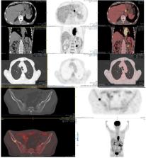
          Precise staging of esophageal cancer (EC) is important for selection of optimal treatment option and prognostication. Aim of this study was to assess the role of 18FDG PET/CT in staging and response evaluation to neoadjuvant chemoradiation (nCR) in EC patients using standardized imaging protocol.

          Material and methods:

          This prospective study was conducted at PET/CT Section of Department of Radiology, Aga Khan University Hospital Karachi, Pakistan from July 2017 till February 2018. We included 34 biopsy proven EC patients who had 18FDG PET/CT and CT of neck, chest and abdomen as part of initial staging. Eleven patients had post-nCR 18FDG PET/CT using standardized imaging protocol as per EANM guidelines. CT and PET/CT based staging was compared. Based on PERCIST criteria, response evaluation was assessed using change in highest SUVmax (%∆SUVmax) in baseline and follow-up scans (primary lesion, node or extra-nodal metastases).

          Results:

          Mean age of cohort was 57 ± 14 years (23 males and 11 females) having adenocarcinoma (AC) in 23 and squamous cell cancer (SCC) in 11 patients. Mean 18FDG dose, uptake time and hepatic SUVmean for baseline scans were 169 ±54 MBq, 65 ±10 minute and 1.91 ± 0.49 which were within ± 10%, ± 15% and ± 20% for follow-up scans in 11 patients respectively. Mean size (craniocaudal dimension in mm) and SUVmax of primary tumor was 56 ±27 mm and 13.4 ± 4.7. Based on 18FDG PET/CT findings, patients were categorized into N0 (10/34), N1 (09/34), N2 (11/34) and N3 (04/34) while 11/32 had stage IV disease. No significant difference was seen in AC and SCC groups. CT found stage IV disease in 3/34 (09%) while PET/CT found in 11/34 (32%; p value: 0.019) cases. PET/CT showed concordance with CT in 41% while discordance (all with upstaging) seen in 59%. On follow-up PET/CT, complete metabolic response was seen in 5/11 (45%) and partial metabolic response was noted in 6/11 (55% - p value non-significant) patients. Median %∆SUVmax over primary lesions was 49.84% (-32.69 -100%) while over nodal sites it was 41.18% (-82.60 -100%).

          Conclusion:

          We conclude that 18FDG PET/CT was found a sensitive tool in initial staging of EC. Compared with CT, it had higher diagnostic accuracy for distant nodal and extra-nodal metastasis. %∆SUVmax between baseline and post-nCR studies acquired with standardized protocol had changed management in more than half of our patients. For response evaluation in EC more studies with standardized 18FDG PET/CT imaging protocols are warranted.

          Related collections

          Most cited references18

          • Record: found
          • Abstract: found
          • Article: not found

          From RECIST to PERCIST: Evolving Considerations for PET response criteria in solid tumors.

          The purpose of this article is to review the status and limitations of anatomic tumor response metrics including the World Health Organization (WHO) criteria, the Response Evaluation Criteria in Solid Tumors (RECIST), and RECIST 1.1. This article also reviews qualitative and quantitative approaches to metabolic tumor response assessment with (18)F-FDG PET and proposes a draft framework for PET Response Criteria in Solid Tumors (PERCIST), version 1.0. PubMed searches, including searches for the terms RECIST, positron, WHO, FDG, cancer (including specific types), treatment response, region of interest, and derivative references, were performed. Abstracts and articles judged most relevant to the goals of this report were reviewed with emphasis on limitations and strengths of the anatomic and PET approaches to treatment response assessment. On the basis of these data and the authors' experience, draft criteria were formulated for PET tumor response to treatment. Approximately 3,000 potentially relevant references were screened. Anatomic imaging alone using standard WHO, RECIST, and RECIST 1.1 criteria is widely applied but still has limitations in response assessments. For example, despite effective treatment, changes in tumor size can be minimal in tumors such as lymphomas, sarcoma, hepatomas, mesothelioma, and gastrointestinal stromal tumor. CT tumor density, contrast enhancement, or MRI characteristics appear more informative than size but are not yet routinely applied. RECIST criteria may show progression of tumor more slowly than WHO criteria. RECIST 1.1 criteria (assessing a maximum of 5 tumor foci, vs. 10 in RECIST) result in a higher complete response rate than the original RECIST criteria, at least in lymph nodes. Variability appears greater in assessing progression than in assessing response. Qualitative and quantitative approaches to (18)F-FDG PET response assessment have been applied and require a consistent PET methodology to allow quantitative assessments. Statistically significant changes in tumor standardized uptake value (SUV) occur in careful test-retest studies of high-SUV tumors, with a change of 20% in SUV of a region 1 cm or larger in diameter; however, medically relevant beneficial changes are often associated with a 30% or greater decline. The more extensive the therapy, the greater the decline in SUV with most effective treatments. Important components of the proposed PERCIST criteria include assessing normal reference tissue values in a 3-cm-diameter region of interest in the liver, using a consistent PET protocol, using a fixed small region of interest about 1 cm(3) in volume (1.2-cm diameter) in the most active region of metabolically active tumors to minimize statistical variability, assessing tumor size, treating SUV lean measurements in the 1 (up to 5 optional) most metabolically active tumor focus as a continuous variable, requiring a 30% decline in SUV for "response," and deferring to RECIST 1.1 in cases that do not have (18)F-FDG avidity or are technically unsuitable. Criteria to define progression of tumor-absent new lesions are uncertain but are proposed. Anatomic imaging alone using standard WHO, RECIST, and RECIST 1.1 criteria have limitations, particularly in assessing the activity of newer cancer therapies that stabilize disease, whereas (18)F-FDG PET appears particularly valuable in such cases. The proposed PERCIST 1.0 criteria should serve as a starting point for use in clinical trials and in structured quantitative clinical reporting. Undoubtedly, subsequent revisions and enhancements will be required as validation studies are undertaken in varying diseases and treatments.
            Bookmark
            • Record: found
            • Abstract: found
            • Article: found
            Is Open Access

            The effect of SUV discretization in quantitative FDG-PET Radiomics: the need for standardized methodology in tumor texture analysis

            FDG-PET-derived textural features describing intra-tumor heterogeneity are increasingly investigated as imaging biomarkers. As part of the process of quantifying heterogeneity, image intensities (SUVs) are typically resampled into a reduced number of discrete bins. We focused on the implications of the manner in which this discretization is implemented. Two methods were evaluated: (1) RD, dividing the SUV range into D equally spaced bins, where the intensity resolution (i.e. bin size) varies per image; and (2) RB, maintaining a constant intensity resolution B. Clinical feasibility was assessed on 35 lung cancer patients, imaged before and in the second week of radiotherapy. Forty-four textural features were determined for different D and B for both imaging time points. Feature values depended on the intensity resolution and out of both assessed methods, RB was shown to allow for a meaningful inter- and intra-patient comparison of feature values. Overall, patients ranked differently according to feature values–which was used as a surrogate for textural feature interpretation–between both discretization methods. Our study shows that the manner of SUV discretization has a crucial effect on the resulting textural features and the interpretation thereof, emphasizing the importance of standardized methodology in tumor texture analysis.
              Bookmark
              • Record: found
              • Abstract: found
              • Article: not found

              Need for standardization of 18F-FDG PET/CT for treatment response assessments.

              Many factors affect standardized uptake values (SUVs) in (18)F-FDG PET/CT. The use of the SUV from a single PET scan in multicenter studies requires the standardization of (18)F-FDG PET/CT procedures. In the context of treatment response assessments (repeated PET scans), many factors may seem to have minor effects on percentage changes in SUVs, provided that imaging procedures are executed in a consistent manner for each subject. However, the use of (18)F-FDG PET/CT in a nonstandardized manner will result in unknown biases and reproducibilities of SUVs and SUV-based response measures. This article provides an overview of the need for standardization in relation to the specific use of SUVs and SUV changes in studies of treatment response assessments.
                Bookmark

                Author and article information

                Journal
                Asian Pac J Cancer Prev
                Asian Pac. J. Cancer Prev
                APJCP
                Asian Pacific Journal of Cancer Prevention : APJCP
                West Asia Organization for Cancer Prevention (Iran )
                1513-7368
                2476-762X
                2019
                : 20
                : 7
                : 2003-2008
                Affiliations
                [1 ] Section of NM and PET/CT imaging, Department of Radiology,
                [3 ] Department of Radiation Oncology, Aga Khan University Hospital (A KUH),
                [2 ] Dow Medical College, Dow University of Health Sciences (DUHS), Karachi, Pakistan.
                Author notes
                [* ]For Correspondence: maseeh.uzzaman@aku.edu
                Article
                10.31557/APJCP.2019.20.7.2003
                6745203
                31350957
                0164455b-2194-4920-bf0b-8dba12fd68c7
                © Asian Pacific Journal of Cancer Prevention

                This work is licensed under the terms of the Creative Commons Attribution-NonCommercial-ShareAlike 4.0 International License ( https://creativecommons.org/licenses/by-nc-sa/4.0/)

                History
                : 6 April 2018
                : 6 June 2019
                Categories
                Research Article

                esophageal cancer- staging- neoadjuvant chemoradiation- 18fdg pet/ct and ct

                Comments

                Comment on this article