5
views
0
recommends
+1 Recommend
0 collections
    0
    shares
      • Record: found
      • Abstract: found
      • Article: found
      Is Open Access

      A Simple and Efficient Method for Visualizing Individual Cells in vivo by Cre-Mediated Single-Cell Labeling by Electroporation (CREMSCLE)

      research-article

      Read this article at

      Bookmark
          There is no author summary for this article yet. Authors can add summaries to their articles on ScienceOpen to make them more accessible to a non-specialist audience.

          Abstract

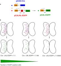
          Efficient methods for visualizing cell morphology in the intact animal are of great benefit to the study of structural development in the nervous system. Quantitative analysis of the complex arborization patterns of brain cells informs cell-type classification, dissection of neuronal circuit wiring, and the elucidation of growth and plasticity mechanisms. Time-lapse single-cell morphological analysis requires labeling and imaging of single cells in situ without contamination from the ramified processes of other nearby cells. Here, using the Xenopus laevis optic tectum as a model system, we describe CRE-Mediated Single-Cell Labeling by Electroporation (CREMSCLE), a technique we developed based on bulk co-electroporation of Cre-dependent inducible expression vectors, together with very low concentrations of plasmid encoding Cre recombinase. This method offers efficient, sparse labeling in any brain area where bulk electroporation is possible. Unlike juxtacellular single-cell electroporation methods, CREMSCLE relies exclusively on the bulk electroporation technique, circumventing the need to precisely position a micropipette next to the target cell. Compared with viral transduction methods, it is fast and safe, generating high levels of expression within 24 h of introducing non-infectious plasmid DNA. In addition to increased efficiency of single-cell labeling, we confirm that CREMSCLE also allows for efficient co-expression of multiple gene products in the same cell. Furthermore, we demonstrate that this method is particularly well-suited for labeling immature neurons to follow their maturation over time. This approach therefore lends itself well to time-lapse morphological studies, particularly in the context of early neuronal development and under conditions that prevent more difficult visualized juxtacellular electroporation.

          Related collections

          Most cited references26

          • Record: found
          • Abstract: found
          • Article: not found

          Biochemistry, mutagenesis, and oligomerization of DsRed, a red fluorescent protein from coral.

          DsRed is a recently cloned 28-kDa fluorescent protein responsible for the red coloration around the oral disk of a coral of the Discosoma genus. DsRed has attracted tremendous interest as a potential expression tracer and fusion partner that would be complementary to the homologous green fluorescent protein from Aequorea, but very little is known of the biochemistry of DsRed. We now show that DsRed has a much higher extinction coefficient and quantum yield than previously reported, plus excellent resistance to pH extremes and photobleaching. In addition, its 583-nm emission maximum can be further shifted to 602 nm by mutation of Lys-83 to Met. However, DsRed has major drawbacks, such as strong oligomerization and slow maturation. Analytical ultracentrifugation proves DsRed to be an obligate tetramer in vitro, and fluorescence resonance energy transfer measurements and yeast two-hybrid assays verify oligomerization in live cells. Also, DsRed takes days to ripen fully from green to red in vitro or in vivo, and mutations such as Lys-83 to Arg prevent the color change. Many potential cell biological applications of DsRed will require suppression of the tetramerization and acceleration of the maturation.
            Bookmark
            • Record: found
            • Abstract: found
            • Article: not found

            Efficient in utero gene transfer system to the developing mouse brain using electroporation: visualization of neuronal migration in the developing cortex.

            We report a novel gene transfer system using electroporation. We used this technique to introduce a marker gene plasmid containing enhanced green fluorescent protein into mouse brains at embryonic day 12-17 without removing the embryos from the uterus. The embryos were allowed to continue to develop in utero, and more than 80% were born normally expressing the exogenous gene. Enhanced green fluorescent protein driven by the cytomegalovirus promoter was strongly expressed in the ventricular zone, radial fibers and migrating neuroblasts, but not in mature neurons, suggesting that the cytomegalovirus promoter is silenced after the cells differentiate into mature neurons. Since there is still no convenient way of visualizing the migrating neuroblasts, especially of distinguishing them from the surrounding mature neurons in the cortical plate, this system should provide a good tool for analysing neuronal migration. In the postnatal lateral cortex, neuroblasts migrated almost "tangentially" along the obliquely running "radial" fibers beneath the cortical plate, and after entering the cortical plate, turned towards the marginal zone and migrated radially. Neurons with primitive dendrites were observed only along the border between the marginal zone and the cortical plate, and never at other sites, such as in the middle of the cortical plate. These results imply that the neuroblasts do terminate migration and start differentiation to mature neurons when they encounter the marginal zone, as has long been suggested. By contrast, when elongation factor 1alpha promoter was used, prominent fluorescence allowed visualization of the entire mature neurons as well. The labeled neurons were observed to send axons to the contralateral cortex where they arborized extensively.Thus, this system is much easier and more efficient than virus-mediated gene transfer, and is useful for gain-of-function analysis of neural cell fate determination, migration, positioning and axon path-finding in mouse embryos.
              Bookmark
              • Record: found
              • Abstract: found
              • Article: not found

              Neocortex patterning by the secreted signaling molecule FGF8.

              A classic model proposes that the mammalian neocortex is divided into areas early in neurogenesis, but the molecular mechanisms that generate the area map have been elusive. Here we provide evidence that FGF8 regulates development of the map from a source in the anterior telencephalon. Using electroporation-mediated gene transfer in mouse embryos, we show that augmenting the endogenous anterior FGF8 signal shifts area boundaries posteriorly, reducing the signal shifts them anteriorly, and introducing a posterior source of FGF8 elicits partial area duplications, revealed by ectopic somatosensory barrel fields. These findings support a role for FGF signaling in specifying positional identity in the neocortex.
                Bookmark

                Author and article information

                Contributors
                Journal
                Front Neural Circuits
                Front Neural Circuits
                Front. Neural Circuits
                Frontiers in Neural Circuits
                Frontiers Media S.A.
                1662-5110
                28 July 2020
                2020
                : 14
                : 47
                Affiliations
                Department of Neurology and Neurosurgery, Montreal Neurological Institute-Hospital, McGill University , Montreal, QC, Canada
                Author notes

                Edited by: Graziella DiCristo, Université de Montréal, Canada

                Reviewed by: Simon Chen, University of Ottawa, Canada; Anthony Holtmaat, Université de Genève, Switzerland

                *Correspondence: Edward S. Ruthazer, edward.ruthazer@ 123456mcgill.ca
                Article
                10.3389/fncir.2020.00047
                7399061
                32848634
                03128eb5-4eda-4980-9d8b-26ec9c27a115
                Copyright © 2020 Schohl, Chorghay and Ruthazer.

                This is an open-access article distributed under the terms of the Creative Commons Attribution License (CC BY). The use, distribution or reproduction in other forums is permitted, provided the original author(s) and the copyright owner(s) are credited and that the original publication in this journal is cited, in accordance with accepted academic practice. No use, distribution or reproduction is permitted which does not comply with these terms.

                History
                : 16 May 2020
                : 08 July 2020
                Page count
                Figures: 6, Tables: 0, Equations: 0, References: 29, Pages: 11, Words: 0
                Funding
                Funded by: Canadian Institutes of Health Research 10.13039/501100000024
                Award ID: FDN-143238
                Categories
                Neuroscience
                Original Research

                Neurosciences
                transfection,morphology,neuron,multiphoton,loxp,xenopus laevis,optic tectum
                Neurosciences
                transfection, morphology, neuron, multiphoton, loxp, xenopus laevis, optic tectum

                Comments

                Comment on this article