9
views
0
recommends
+1 Recommend
0 collections
    0
    shares
      • Record: found
      • Abstract: found
      • Article: found
      Is Open Access

      Dengue fatal cases present virus-specific HMGB1 response in peripheral organs

      research-article

      Read this article at

      Bookmark
          There is no author summary for this article yet. Authors can add summaries to their articles on ScienceOpen to make them more accessible to a non-specialist audience.

          Abstract

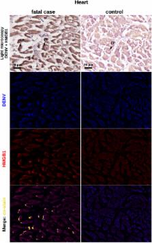
          Dengue is an important infectious disease that presents high incidence and yields a relevant number of fatal cases (about 20,000) every year worldwide. Despite its epidemiological relevance, there are many knowledge gaps concerning dengue pathogenesis, especially with regards to the circumstances that drive a mild clinical course to a severe disease. In this work, we investigated the participation of high mobility group box 1 (HMGB1), an important modulator of inflammation, in dengue fatal cases. Histopathological and ultrastructural analyses revealed that liver, lung and heart post-mortem samples were marked by tissue abnormalities, such as necrosis and apoptotic cell death. These observations go in line with an HMGB1-mediated response and raised concerns regarding the participation of this cytokine in promoting/perpetuating inflammation in severe dengue. Further experiments of immunohistochemistry (IHC) showed increased expression of cytoplasmic HMGB1 in dengue-extracted tissues when compared to non-dengue controls. Co-staining of DENV RNA and HMGB1 in the host cell cytoplasm, as found by in situ hybridization and IHC, confirmed the virus specific induction of the HMGB1-mediated response in these peripheral tissues. This report brings the first in-situ evidence of the participation of HMGB1 in severe dengue and highlights novel considerations in the development of dengue immunopathogenesis.

          Related collections

          Most cited references55

          • Record: found
          • Abstract: found
          • Article: not found

          The extracellular release of HMGB1 during apoptotic cell death.

          High mobility group box 1 protein (HMGB1) is a non-histone nuclear protein with dual function. Inside the cell, HMGB1 binds DNA and regulates transcription, whereas outside the cell, it serves as a cytokine and mediates the late effects of LPS. The movement of HMGB1 into the extracellular space has been demonstrated for macrophages stimulated with LPS as well as cells undergoing necrosis but not apoptosis. The differential release of HMGB1 during death processes could reflect the structure of chromatin in these settings as well as the mechanisms for HMGB1 translocation. Since apoptotic cells can release some nuclear molecules such as DNA to which HMGB1 can bind, we therefore investigated whether HMGB1 release can occur during apoptosis as well as necrosis. For this purpose, Jurkat cells were treated with chemical inducers of apoptosis (staurosporine, etoposide, or camptothecin), and HMGB1 release into the medium was assessed by Western blotting. Results of these experiments indicate that HMGB1 appears in the media of apoptotic Jurkat cells in a time-dependent manner and that this release can be reduced by Z-VAD-fmk. Panc-1 and U937 cells treated with these agents showed similar release. In addition, HeLa cells induced to undergo apoptosis showed HMGB1 release. Furthermore, we showed using confocal microscopy that HMGB1 and DNA change their nuclear location in Jurkat cells undergoing apoptosis. Together, these studies indicate that HMGB1 release can occur during the course of apoptosis as well as necrosis and suggest that the release process may vary with cell type.
            Bookmark
            • Record: found
            • Abstract: found
            • Article: not found

            HMGB1 is an endogenous immune adjuvant released by necrotic cells.

            Immune responses against pathogens require that microbial components promote the activation of antigen-presenting cells (APCs). Autoimmune diseases and graft rejections occur in the absence of pathogens; in these conditions, endogenous molecules, the so-called 'innate adjuvants', activate APCs. Necrotic cells contain and release innate adjuvants; necrotic cells also release high-mobility group B1 protein (HMGB1), an abundant and conserved constituent of vertebrate nuclei. Here, we show that necrotic HMGB1(-/-) cells have a reduced ability to activate APCs, and HMGB1 blockade reduces the activation induced by necrotic wild-type cell supernatants. In vivo, HMGB1 enhances the primary antibody responses to soluble antigens and transforms poorly immunogenic apoptotic lymphoma cells into efficient vaccines.
              Bookmark
              • Record: found
              • Abstract: found
              • Article: found
              Is Open Access

              Dengue Virus NS1 Disrupts the Endothelial Glycocalyx, Leading to Hyperpermeability

              Dengue is the most prevalent arboviral disease in humans and a major public health problem worldwide. Systemic plasma leakage, leading to hypovolemic shock and potentially fatal complications, is a critical determinant of dengue severity. Recently, we and others described a novel pathogenic effect of secreted dengue virus (DENV) non-structural protein 1 (NS1) in triggering hyperpermeability of human endothelial cells in vitro and systemic vascular leakage in vivo. NS1 was shown to activate toll-like receptor 4 signaling in primary human myeloid cells, leading to secretion of pro-inflammatory cytokines and vascular leakage. However, distinct endothelial cell-intrinsic mechanisms of NS1-induced hyperpermeability remained to be defined. The endothelial glycocalyx layer (EGL) is a network of membrane-bound proteoglycans and glycoproteins lining the vascular endothelium that plays a key role in regulating endothelial barrier function. Here, we demonstrate that DENV NS1 disrupts the EGL on human pulmonary microvascular endothelial cells, inducing degradation of sialic acid and shedding of heparan sulfate proteoglycans. This effect is mediated by NS1-induced expression of sialidases and heparanase, respectively. NS1 also activates cathepsin L, a lysosomal cysteine proteinase, in endothelial cells, which activates heparanase via enzymatic cleavage. Specific inhibitors of sialidases, heparanase, and cathepsin L prevent DENV NS1-induced EGL disruption and endothelial hyperpermeability. All of these effects are specific to NS1 from DENV1-4 and are not induced by NS1 from West Nile virus, a related flavivirus. Together, our data suggest an important role for EGL disruption in DENV NS1-mediated endothelial dysfunction during severe dengue disease.
                Bookmark

                Author and article information

                Contributors
                edsonrao@gmail.com
                marciano@ioc.fiocruz.br
                Journal
                Sci Rep
                Sci Rep
                Scientific Reports
                Nature Publishing Group UK (London )
                2045-2322
                22 November 2017
                22 November 2017
                2017
                : 7
                : 16011
                Affiliations
                [1 ]ISNI 0000 0001 2294 473X, GRID grid.8536.8, Laboratóio de Modelagem Molecular, Instituto de Química Orgânica, Universidade Federal do Rio de Janeiro, ; Rio de Janeiro, Brazil
                [2 ]Instituto de Criminalística, Tocantins, Brazil
                [3 ]ISNI 0000 0001 2285 7943, GRID grid.261331.4, Ohio State University Comprehensive Cancer Center, Columbus, ; Ohio, United States of America
                [4 ]GRID grid.430298.4, Phylogeny Inc, Powell, ; Ohio, United States of America
                [5 ]ISNI 0000 0001 2294 473X, GRID grid.8536.8, Laboratório de Genômica Estrutural, Instituto de Biofísica Carlos Chagas Filho, Universidade Federal do Rio de Janeiro, ; Rio de Janeiro, Brazil
                [6 ]ISNI 0000 0001 0723 0931, GRID grid.418068.3, Laboratório Interdisciplinar de Pesquisas Médicas, Instituto Oswaldo Cruz, Fundação Oswaldo Cruz, Rio de Janeiro, ; RJ, Brazil
                [7 ]ISNI 0000 0001 2237 7915, GRID grid.467095.9, Anatomia Patológica, Hospital Gaffrée Guinle, Universidade Federal do Estado do Rio de Janeiro, ; Rio de Janeiro, Brazil
                [8 ]ISNI 0000 0001 2294 473X, GRID grid.8536.8, Laboratório de Biologia das Células Gliais, Instituto de Ciências Biomédicas, Universidade Federal do Rio de Janeiro, ; Rio de Janeiro, Brazil
                Author information
                http://orcid.org/0000-0002-8606-7454
                http://orcid.org/0000-0002-4452-9542
                Article
                16197
                10.1038/s41598-017-16197-5
                5700165
                29167501
                051a1a32-cbd4-4533-9093-3948025f0167
                © The Author(s) 2017

                Open Access This article is licensed under a Creative Commons Attribution 4.0 International License, which permits use, sharing, adaptation, distribution and reproduction in any medium or format, as long as you give appropriate credit to the original author(s) and the source, provide a link to the Creative Commons license, and indicate if changes were made. The images or other third party material in this article are included in the article’s Creative Commons license, unless indicated otherwise in a credit line to the material. If material is not included in the article’s Creative Commons license and your intended use is not permitted by statutory regulation or exceeds the permitted use, you will need to obtain permission directly from the copyright holder. To view a copy of this license, visit http://creativecommons.org/licenses/by/4.0/.

                History
                : 2 October 2017
                : 8 November 2017
                Categories
                Article
                Custom metadata
                © The Author(s) 2017

                Uncategorized
                Uncategorized

                Comments

                Comment on this article