2
views
0
recommends
+1 Recommend
0 collections
    0
    shares
      • Record: found
      • Abstract: found
      • Article: found
      Is Open Access

      LKB1 suppresses androgen synthesis in a mouse model of hyperandrogenism via IGF‐1 signaling

      research-article

      Read this article at

      Bookmark
          There is no author summary for this article yet. Authors can add summaries to their articles on ScienceOpen to make them more accessible to a non-specialist audience.

          Abstract

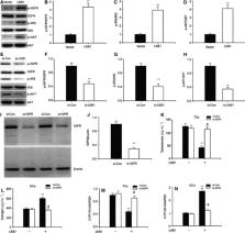
          Polycystic ovary syndrome ( PCOS) is a major cause of anovulatory sterility in women, and most PCOS patients exhibit hyperandrogenism ( HA). Liver kinase b1 ( LKB1) is a tumor suppressor that has recently been reported to be involved in PCOS. However, the mechanism by which LKB1 affects HA has not previously been elucidated. We report here that ovarian LKB1 levels are significantly decreased in a female mouse model of HA. Moreover, we report that LKB1 expression is inhibited by elevated androgens via activation of androgen receptors. In addition, LKB1 treatment was observed to suppress androgen synthesis in theca cells and promote estrogen production in granulosa cells by regulating steroidogenic enzyme expression. As expected, LKB1 knockdown inhibited estrogen levels and enhanced androgen levels, and LKB1‐transgenic mice were protected against HA. The effect of LKB1 appears to be mediated via IGF‐1 signaling. In summary, we describe here a key role for LKB1 in controlling sex hormone levels.

          Related collections

          Most cited references36

          • Record: found
          • Abstract: found
          • Article: not found

          Development of polycystic ovary syndrome: involvement of genetic and environmental factors.

          We have recently proposed that polycystic ovary syndrome (PCOS) has its origin in fetal life. This hypothesis is based on data from animal models (rhesus monkey or sheep that have been exposed prenatally to high doses of androgen) and is supported by clinical studies. It is suggested that, in human females, exposure to excess androgen, at any stage from fetal development of the ovary to the onset of puberty, leads to many of the characteristic features of PCOS, including abnormalities of luteinizing hormone secretion and insulin resistance. It is likely that, in humans with PCOS, the development of the PCOS phenotype results primarily from a genetic predisposition for the fetal ovary to hypersecrete androgen. At present, it is unclear whether the maternal environment directly influences the development of PCOS in the offspring. Maternal androgen excess is unlikely to affect the fetus, because the placenta presents an effective barrier, but metabolic disturbances during pregnancy could affect development of the syndrome in the fetus. In postnatal life, the natural history of PCOS can be further modified by factors affecting insulin secretion and/or action, most importantly, nutrition. We now have evidence for a disorder of early follicular development in the polycystic ovary that is consistent with an increased population of primordial follicles in the fetal ovary. It remains to be determined whether this phenomenon is the cause or the effect of increased exposure to androgen within the ovary. PCOS is the commonest endocrine disorder in women. It is not only a very prevalent cause of anovulatory infertility, menstrual disturbances and hirsutism, but it is also a major risk factor for the development of type 2 diabetes mellitus in later life. The aetiology of the syndrome remains uncertain but there is increasing evidence for a genetic basis. PCOS very often becomes clinically manifest during adolescence with maturation of the hypothalamic-pituitary-ovarian axis but the genesis of the syndrome may be during very early development - perhaps even in utero. In this review, this hypothesis is explored in the light of clinical, biochemical and genetic research.
            Bookmark
            • Record: found
            • Abstract: found
            • Article: found
            Is Open Access

            FOXO3 Is a Glucocorticoid Receptor Target and Regulates LKB1 and Its Own Expression Based on Cellular AMP Levels via a Positive Autoregulatory Loop

            FOXO3 is a transcription factor involved in the regulation of multiple physiological processes including cell cycle arrest, apoptosis, oxidative stress-response and energy metabolism. Although much is known about its post-translational modification, the transcriptional regulation of FOXO3, as well as the cross-talk between transcription and post-translational events, is still poorly understood. In the present study, we show that FOXO3 is an immediate early glucocorticoid receptor (GR) target, whose transcription is even further enhanced by conditions that mimic metabolic stress. Induction of FOXO3 transcription by GR-binding steroids was reversed by concomitant treatment with the GR antagonist RU-486, but further enhanced by stimuli that activate the AMP-activated protein kinase (AMPK). Analysis of genomic DNA and chromatin immunoprecipitation, as well as luciferase reporter assays, revealed two functional glucocorticoid responsive elements within the FOXO3 promoter. Furthermore, we provide functional evidence for a phosphorylation switch that explains how glucocorticoids induce transcriptional activation of the gene but subsequently inactivate the corresponding protein by site-specific phosphorylation. Only when AMPK is stimulated, pre-existing FOXO3 becomes reverted toward an active form. Energy deprived conditions thus activate FOXO3 on two different levels, namely transcriptional and post-translational. In that way, FOXO3 acts as a metabolic stress sensor that coordinates expression of LKB1, the master upstream kinase involved in metabolic sensing, depending on the energy status of the cell. Additionally, we show that FOXO3 binds and activates its own promoter via a positive autoregulatory feedback loop. In conclusion, our data explain how catabolic glucocorticoid hormones and high intracellular AMP levels cooperate in inducing FOXO3 transcription and in activating the corresponding protein.
              Bookmark
              • Record: found
              • Abstract: found
              • Article: not found

              LKB1 promotes metabolic flexibility in response to energy stress

              The Liver Kinase B1 (LKB1) tumor suppressor acts as a metabolic energy sensor to regulate AMP-activated protein kinase (AMPK) signaling and is commonly mutated in various cancers, including non-small cell lung cancer (NSCLC). Tumor cells deficient in LKB1 may be uniquely sensitized to metabolic stresses, which may offer a therapeutic window in oncology. To address this question we have explored how functional LKB1 impacts the metabolism of NSCLC cells using 13 C metabolic flux analysis. Isogenic NSCLC cells expressing functional LKB1 exhibited higher flux through oxidative mitochondrial pathways compared to those deficient in LKB1. Re-expression of LKB1 also increased the capacity of cells to oxidize major mitochondrial substrates, including pyruvate, fatty acids, and glutamine. Furthermore, LKB1 expression promoted an adaptive response to energy stress induced by anchorage-independent growth. Finally, this diminished adaptability sensitized LKB1-deficient cells to combinatorial inhibition of mitochondrial complex I and glutaminase. Together, our data implicate LKB1 as a major regulator of adaptive metabolic reprogramming and suggest synergistic pharmacological strategies for mitigating LKB1-deficient NSCLC tumor growth.
                Bookmark

                Author and article information

                Contributors
                yingxu5434664@163.com
                Journal
                FEBS Open Bio
                FEBS Open Bio
                10.1002/(ISSN)2211-5463
                FEB4
                FEBS Open Bio
                John Wiley and Sons Inc. (Hoboken )
                2211-5463
                12 September 2019
                October 2019
                : 9
                : 10 ( doiID: 10.1002/feb4.v9.10 )
                : 1817-1825
                Affiliations
                [ 1 ] Department of Obstetrics and Gynecology the 476th Hospital of PLA Fuzhou China
                [ 2 ] Fuzong Clinical College Fujian Medical University Fuzhou China
                [ 3 ] Department of Obstetrics and Gynecology Zhongda Hospital Southeast University Nanjing China
                [ 4 ] Key Laboratory of OptoElectronic Science and Technology for Medicine of Ministry of Education Fujian Provincial Key Laboratory of Photonics Technology Fujian Normal University Fuzhou China
                Author notes
                [*] [* ] Correspondence

                Y. Xu, Department of Obstetrics and Gynecology, the 476th Hospital of PLA, No. 90 Guo Cuo Li, Hong Tang Road, Cangshan District, Fuzhou, Fujian 350002, China

                Tel: +86 591 88611331

                E‐mail: yingxu5434664@ 123456163.com

                Article
                FEB412723
                10.1002/2211-5463.12723
                6768104
                31433577
                06d00365-b8d5-46af-a28f-654822fc8188
                © 2019 The Authors. Published by FEBS Press and John Wiley & Sons Ltd.

                This is an open access article under the terms of the http://creativecommons.org/licenses/by/4.0/ License, which permits use, distribution and reproduction in any medium, provided the original work is properly cited.

                History
                : 30 January 2019
                : 30 May 2019
                : 19 August 2019
                Page count
                Figures: 4, Tables: 0, Pages: 9, Words: 4655
                Categories
                Research Article
                Research Articles
                Custom metadata
                2.0
                feb412723
                October 2019
                Converter:WILEY_ML3GV2_TO_NLMPMC version:5.6.9 mode:remove_FC converted:30.09.2019

                hyperandrogenism,igf‐1 signal,lkb1,polycystic ovary syndrome

                Comments

                Comment on this article