3
views
0
recommends
+1 Recommend
0 collections
    0
    shares
      • Record: found
      • Abstract: found
      • Article: found
      Is Open Access

      Continuous reduction in cerebral oxygenation during endurance exercise in patients with pulmonary arterial hypertension

      research-article

      Read this article at

      Bookmark
          There is no author summary for this article yet. Authors can add summaries to their articles on ScienceOpen to make them more accessible to a non-specialist audience.

          Abstract

          Background

          Patients with pulmonary arterial hypertension (PAH) have lower cerebral blood flow (CBF) and oxygenation compared to healthy sedentary subjects, the latter negatively correlating with exercise capacity during incremental cycling exercise. We hypothesized that patients would also exhibit altered CBF and oxygenation during endurance exercise, which would correlate with endurance time.

          Methods

          Resting and exercise cardiorespiratory parameters, blood velocity in the middle cerebral artery (MCAv; transcranial doppler) and cerebral oxygenation (relative changes in cerebral tissue oxygenation index (ΔcTOI) and cerebral deoxyhemoglobin (ΔcHHb); near‐infrared spectroscopy) were continuously monitored in nine PAH patients and 10 healthy‐matched controls throughout endurance exercise. Cardiac output (CO), systemic blood pressure (BP) and oxygen saturation (SpO 2), ventilatory metrics and end‐tidal CO 2 pressure (P ETCO 2) were also assessed noninvasively.

          Results

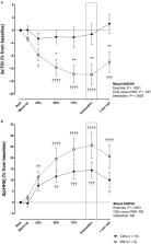
          Despite a lower workload and endurance oxygen consumption, similar CO and systemic BP, ΔcTOI was lower in PAH patients compared to controls ( p < .01 for interaction). As expected during exercise, patients were characterized by an altered MCAv response to exercise, a lower P ETCO 2 and SpO 2, as wells as a higher minute‐ventilation/CO 2 production ratio ( V ˙ E / V ˙ CO 2 ratio). An uncoupling between changes in MCAv and P ETCO 2 during the cycling endurance exercise was also progressively apparent in PAH patients, but absent in healthy controls. Both cHHb and ΔcTOI correlated with V ˙ E / V ˙ CO 2 ratio ( r = 0.50 and r = −0.52; both p < .05 respectively), but not with endurance time.

          Conclusion

          PAH patients present an abnormal cerebrovascular profile during endurance exercise with a lower cerebral oxygenation that correlate with hyperventilation but not endurance exercise time. These findings complement the physiological characterization of the cerebral vascular responses to exercise in PAH patients.

          Abstract

          The novel findings of this study are that abnormal cerebrovascular responses to exercise are present during an endurance exercise protocol in patients with pulmonary arterial hypertension. It includes a reduction in cerebral oxygenation associated with higher minute‐ventilation/carbon dioxide production rather than endurance time, and an uncoupling between changes in mean blood velocity in the middle cerebral artery and end‐tidal carbon dioxide partial pressure during exercise in patients only. These findings add to the existing literature reporting abnormalities in cerebral blood flow determinants in pulmonary arterial hypertension.

          Related collections

          Most cited references47

          • Record: found
          • Abstract: found
          • Article: not found

          Pericyte degeneration leads to neurovascular uncoupling and limits oxygen supply to brain

          Pericytes are perivascular mural cells of brain capillaries that are positioned centrally within the neurovascular unit between endothelial cells, astrocytes and neurons. This unique position allows them to play a major role in regulating key neurovascular functions of the brain. The role of pericytes in the regulation of cerebral blood flow (CBF) and neurovascular coupling remains, however, debatable. Using loss-of-function pericyte-deficient mice, here we show that pericyte degeneration diminishes global and individual capillary CBF responses to neuronal stimulus resulting in neurovascular uncoupling, reduced oxygen supply to brain and metabolic stress. We show that these neurovascular deficits lead over time to impaired neuronal excitability and neurodegenerative changes. Thus, pericyte degeneration as seen in neurological disorders such as Alzheimer’s disease may contribute to neurovascular dysfunction and neurodegeneration associated with human disease.
            Bookmark
            • Record: found
            • Abstract: found
            • Article: not found

            Utility of transcranial Doppler ultrasound for the integrative assessment of cerebrovascular function.

            There is considerable utility in the use of transcranial Doppler ultrasound (TCD) to assess cerebrovascular function. The brain is unique in its high energy and oxygen demand but limited capacity for energy storage that necessitates an effective means of regional blood delivery. The relative low cost, ease-of-use, non-invasiveness, and excellent temporal resolution of TCD make it an ideal tool for the examination of cerebrovascular function in both research and clinical settings. TCD is an efficient tool to access blood velocities within the cerebral vessels, cerebral autoregulation, cerebrovascular reactivity to CO(2), and neurovascular coupling, in both physiological states and in pathological conditions such as stroke and head trauma. In this review, we provide: (1) an overview of TCD methodology with respect to other techniques; (2) a methodological synopsis of the cerebrovascular exam using TCD; (3) an overview of the physiological mechanisms involved in regulation of the cerebral blood flow; (4) the utility of TCD for assessment of cerebrovascular pathology; and (5) recommendations for the assessment of four critical and complimentary aspects of cerebrovascular function: intra-cranial blood flow velocity, cerebral autoregulation, cerebral reactivity, and neurovascular coupling. The integration of these regulatory mechanisms from an integrated systems perspective is discussed, and future research directions are explored. Copyright © 2011 Elsevier B.V. All rights reserved.
              Bookmark
              • Record: found
              • Abstract: found
              • Article: not found

              Cerebral blood flow during exercise: mechanisms of regulation.

              The response of cerebral vasculature to exercise is different from other peripheral vasculature; it has a small vascular bed and is strongly regulated by cerebral autoregulation and the partial pressure of arterial carbon dioxide (Pa(CO(2))). In contrast to other organs, the traditional thinking is that total cerebral blood flow (CBF) remains relatively constant and is largely unaffected by a variety of conditions, including those imposed during exercise. Recent research, however, indicates that cerebral neuronal activity and metabolism drive an increase in CBF during exercise. Increases in exercise intensity up to approximately 60% of maximal oxygen uptake produce elevations in CBF, after which CBF decreases toward baseline values because of lower Pa(CO(2)) via hyperventilation-induced cerebral vasoconstriction. This finding indicates that, during heavy exercise, CBF decreases despite the cerebral metabolic demand. In contrast, this reduced CBF during heavy exercise lowers cerebral oxygenation and therefore may act as an independent influence on central fatigue. In this review, we highlight methodological considerations relevant for the assessment of CBF and then summarize the integrative mechanisms underlying the regulation of CBF at rest and during exercise. In addition, we examine how CBF regulation during exercise is altered by exercise training, hypoxia, and aging and suggest avenues for future research.
                Bookmark

                Author and article information

                Contributors
                simon.malenfant.2@ulaval.ca
                Journal
                Physiol Rep
                Physiol Rep
                10.1002/(ISSN)2051-817X
                PHY2
                physreports
                Physiological Reports
                John Wiley and Sons Inc. (Hoboken )
                2051-817X
                18 March 2020
                March 2020
                : 8
                : 6 ( doiID: 10.14814/phy2.v8.6 )
                : e14389
                Affiliations
                [ 1 ] Pulmonary Hypertension and Vascular Biology Research Group Quebec City QC Canada
                [ 2 ] Quebec Heart and Lung Institute Research Center Université Laval Quebec City QC Canada
                [ 3 ] Department of Medicine Faculty of Medicine Université Laval Quebec City QC Canada
                [ 4 ] Department of Kinesiology Faculty of Medicine Université Laval Quebec City QC Canada
                Author notes
                [*] [* ] Correspondence

                Simon Malenfant, Pulmonary Hypertension Research Group of the Quebec Heart and Lung Institute Research Center, Laval University, Quebec City, QC G1V 4G5, Canada.

                Email: simon.malenfant.2@ 123456ulaval.ca

                Author information
                https://orcid.org/0000-0002-3911-3702
                https://orcid.org/0000-0002-6254-5044
                Article
                PHY214389
                10.14814/phy2.14389
                7080869
                32189447
                06f21d01-d674-4269-8cf0-2bfbfcc974fa
                © 2020 The Authors. Physiological Reports published by Wiley Periodicals, Inc. on behalf of The Physiological Society and the American Physiological Society.

                This is an open access article under the terms of the http://creativecommons.org/licenses/by/4.0/ License, which permits use, distribution and reproduction in any medium, provided the original work is properly cited.

                History
                : 07 February 2020
                : 13 February 2020
                Page count
                Figures: 2, Tables: 3, Pages: 12, Words: 7970
                Funding
                Funded by: Fond de Recherche du Québec – Santé
                Award ID: 27818
                Funded by: Postdoctoral scholarship from the Canadian Institutes of Health Research
                Award ID: 358583
                Categories
                Cardiovascular Physiology
                Respiratory Conditions Disorder and Diseases
                Central Nervous System
                Endurance and Performance
                Original Research
                Original Research
                Custom metadata
                2.0
                March 2020
                Converter:WILEY_ML3GV2_TO_JATSPMC version:5.7.8 mode:remove_FC converted:19.03.2020

                cerebral blood flow,cerebral oxygenation,cycling endurance test,exercise physiology,pulmonary arterial hypertension,pulmonary hypertension

                Comments

                Comment on this article