37
views
0
recommends
+1 Recommend
0 collections
    8
    shares
      • Record: found
      • Abstract: found
      • Article: found
      Is Open Access

      Nuclear localization and cytosolic overexpression of LASP-1 correlates with tumor size and nodal-positivity of human breast carcinoma

      research-article

      Read this article at

      Bookmark
          There is no author summary for this article yet. Authors can add summaries to their articles on ScienceOpen to make them more accessible to a non-specialist audience.

          Abstract

          Background

          LIM and SH3 protein 1 (LASP-1), initially identified from human breast cancer, is a specific focal adhesion protein involved in cell proliferation and migration, which was reported to be overexpressed in 8–12 % of human breast cancers and thought to be exclusively located in cytoplasm.

          Methods

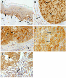
          In the present work we analyzed the cellular and histological expression pattern of LASP-1 and its involvement in biological behavior of human breast cancer through correlation with standard clinicopathological parameters and expression of c-erbB2 (HER-2/neu), estrogen- (ER) and progesterone-receptors (PR). For this purpose immunohistochemical staining intensity and percentage of stained cells were semi-quantitatively rated to define a LASP-1 immunoreactive score (LASP-1-IRS). LASP-1-IRS was determined in 83 cases of invasive ductal breast carcinomas, 25 ductal carcinomas in situ (DCIS) and 18 fibroadenomas. Cellular LASP-1 distribution and expression pattern was visualized by immunofluorescence and confocal microscopy and assessed through separate Western blots of nuclear and cytosol preparations of BT-20, MCF-7, MDA-MB231, and ZR-75/1 breast cancer cells.

          Results

          Statistical analysis revealed that the resulting LASP-1-IRS was significantly higher in invasive carcinomas compared to fibroadenomas (p = 0.0176). Strong cytoplasmatic expression of LASP-1 was detected in 55.4 % of the invasive carcinomas, which correlated significantly with nuclear LASP-1-positivity (p = 0.0014), increased tumor size (p = 0.0159) and rate of nodal-positivity (p = 0.0066). However, levels of LASP-1 expression did not correlate with average age at time point of diagnosis, histological tumor grading, c-erbB2-, ER- or PR-expression.

          Increased nuclear localization and cytosolic expression of LASP-1 was found in breast cancer with higher tumor stage as well as in rapidly proliferating epidermal basal cells. Confocal microscopy and separate Western blots of cytosolic and nuclear preparations confirmed nuclear localization of LASP-1.

          Conclusion

          The current data provide evidence that LASP-1 is not exclusively a cytosolic protein, but is also detectable within the nucleus. Increased expression of LASP-1 in vivo is present in breast carcinomas with higher tumor stage and therefore may be related with worse prognosis concerning patients' overall survival.

          Related collections

          Most cited references38

          • Record: found
          • Abstract: not found
          • Article: not found

          [Recommendation for uniform definition of an immunoreactive score (IRS) for immunohistochemical estrogen receptor detection (ER-ICA) in breast cancer tissue].

            Bookmark
            • Record: found
            • Abstract: not found
            • Article: not found

            Tamoxifen in the treatment of breast cancer.

              Bookmark
              • Record: found
              • Abstract: found
              • Article: not found

              Relevance of breast cancer cell lines as models for breast tumours: an update.

              The number of available breast cancer cell (BCC) lines is small, and only a very few of them have been extensively studied. Whether they are representative of the tumours from which they originated remains a matter of debate. Whether their diversity mirrors the well-known inter-tumoural heterogeneity is another essential question. While numerous similarities have long been found between cell lines and tumours, recent technical advances, including the use of micro-arrays and comparative genetic analysis, have brought new data to the discussion. This paper presents most of the BCC lines that have been described in some detail to date. It evaluates the accuracy of the few of them widely used (MCF-7, T-47D, BT-474, SK-BR-3, MDA-MB-231, Hs578T) as tumour models. It is concluded that BCC lines are likely to reflect, to a large extent, the features of cancer cells in vivo. The importance of oestrogen receptor-alpha (gene ESR1 ) and Her-2/ neu ( ERBB2 ) as classifiers for cell lines and tumours is underlined. The recourse to a larger set of cell lines is suggested since the exact origin of some of the widely used lines remains ambiguous. Investigations on additional specific lines are expected to improve our knowledge of BCC and of the dialogue that these maintain with their surrounding normal cells in vivo.
                Bookmark

                Author and article information

                Journal
                BMC Cancer
                BMC Cancer
                BioMed Central
                1471-2407
                2007
                23 October 2007
                : 7
                : 198
                Affiliations
                [1 ]Institute of Clinical Biochemistry and Pathobiochemistry, University of Wuerzburg, Grombuehlstr. 12, D-97080 Wuerzburg, Germany
                [2 ]Department of Obstetrics and Gynecology, University of Wuerzburg, Josef-Schneider-Str. 4, D-97080 Wuerzburg, Germany
                [3 ]Institute of Pathology, University of Wuerzburg, Josef-Schneider-Str. 2, D-97080 Wuerzburg, Germany
                Article
                1471-2407-7-198
                10.1186/1471-2407-7-198
                2151952
                17956604
                07a3f2fe-f649-40ad-a617-964e93d2ca0f
                Copyright © 2007 Grunewald et al; licensee BioMed Central Ltd.

                This is an Open Access article distributed under the terms of the Creative Commons Attribution License ( http://creativecommons.org/licenses/by/2.0), which permits unrestricted use, distribution, and reproduction in any medium, provided the original work is properly cited.

                History
                : 13 July 2007
                : 23 October 2007
                Categories
                Research Article

                Oncology & Radiotherapy
                Oncology & Radiotherapy

                Comments

                Comment on this article