2
views
0
recommends
+1 Recommend
0 collections
    0
    shares
      • Record: found
      • Abstract: found
      • Article: found
      Is Open Access

      Loss of CDKN1A mRNA and Protein Expression Are Independent Predictors of Poor Outcome in Chromophobe Renal Cell Carcinoma Patients

      research-article

      Read this article at

      Bookmark
          There is no author summary for this article yet. Authors can add summaries to their articles on ScienceOpen to make them more accessible to a non-specialist audience.

          Abstract

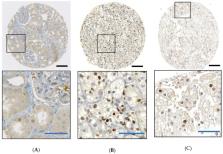
          Chromophobe renal cell carcinoma (chRCC) patients have good prognosis. Only 5%–10% patients die of metastatic disease after tumorectomy, but tumor progression cannot be predicted by histopathological parameters alone. chRCC are characterized by losses of many chromosomes, whereas gene mutations are rare. In this study, we aim at identifying genes indicating chRCC progression. A bioinformatic approach was used to correlate chromosomal loss and mRNA expression from 15287 genes from The Cancer Genome Atlas (TCGA) database. All genes in TCGA chromophobe renal cancer dataset (KICH) for which a significant correlation between chromosomal loss and mRNA expression was shown, were identified and their associations with outcome was assessed. Genome-wide DNA copy-number alterations were analyzed by Affymetrix OncoScan ® CNV FFPE Microarrays in a second cohort of Swiss chRCC. In both cohorts, tumors with loss of chromosomes 2, 6, 10, 13, 17 and 21 had signs of tumor progression. There were 4654 genes located on these chromosomes, and 13 of these genes had reduced mRNA levels, which was associated with poor outcome in chRCC. Decreased CDKN1A expression at mRNA ( p = 0.02) and protein levels ( p = 0.02) were associated with short overall survival and were independent predictors of prognosis ( p < 0.01 and <0.05 respectively). CDKN1A expression status is a prognostic biomarker independent of tumor stage. CDKN1A immunohistochemistry may be used to identify chRCC patients at greater risk of disease progression.

          Related collections

          Most cited references55

          • Record: found
          • Abstract: found
          • Article: not found

          The somatic genomic landscape of chromophobe renal cell carcinoma.

          We describe the landscape of somatic genomic alterations of 66 chromophobe renal cell carcinomas (ChRCCs) on the basis of multidimensional and comprehensive characterization, including mtDNA and whole-genome sequencing. The result is consistent that ChRCC originates from the distal nephron compared with other kidney cancers with more proximal origins. Combined mtDNA and gene expression analysis implicates changes in mitochondrial function as a component of the disease biology, while suggesting alternative roles for mtDNA mutations in cancers relying on oxidative phosphorylation. Genomic rearrangements lead to recurrent structural breakpoints within TERT promoter region, which correlates with highly elevated TERT expression and manifestation of kataegis, representing a mechanism of TERT upregulation in cancer distinct from previously observed amplifications and point mutations.
            Bookmark
            • Record: found
            • Abstract: found
            • Article: not found

            Comparisons of outcome and prognostic features among histologic subtypes of renal cell carcinoma.

            Our objective was to compare cancer-specific survival and to examine associations with outcome among the histologic subtypes of renal cell carcinoma (RCC). We studied 2385 patients whose first surgery between 1970 and 2000 was a radical nephrectomy for sporadic, unilateral RCC. All RCC tumors were classified following the 1997 Union Internationale Contre le Cancer and American Joint Committee on Cancer guidelines. There were 1985 (83.2%) patients with clear cell, 270 (11.3%) with papillary, 102 (4.3%) with chromophobe, 6 (0.3%) with collecting duct, 5 (0.3%) with purely sarcomatoid RCC and no underlying histologic subtype, and 17 (0.7%) with RCC, not otherwise specified. Cancer-specific survival rates at 5 years for patients with clear cell, papillary, and chromophobe RCC were 68.9%, 87.4%, and 86.7%, respectively. Patients with clear cell RCC had a poorer prognosis compared with patients with papillary and chromophobe RCC (p <0.001). This difference in outcome was observed even after stratifying by 1997 tumor stage and nuclear grade. There was no significant difference in cancer-specific survival between patients with papillary and chromophobe RCC (p = 0.918). The 1997 TNM stage, tumor size, presence of a sarcomatoid component, and nuclear grade were significantly associated with death from clear cell, papillary, and chromophobe RCC. Histologic tumor necrosis was significantly associated with death from clear cell and chromophobe RCC, but not with death from papillary RCC. Our results demonstrate that there are significant differences in outcome and associations with outcome for the different histologic subtypes of RCC, highlighting the need for accurate subtyping.
              Bookmark
              • Record: found
              • Abstract: found
              • Article: not found

              Cytoplasmic localization of p21Cip1/WAF1 by Akt-induced phosphorylation in HER-2/neu-overexpressing cells.

              Amplification or overexpression of HER-2/neu in cancer cells confers resistance to apoptosis and promotes cell growth. The cellular localization of p21Cip1/WAF1 has been proposed to be critical either in promoting cell survival or in inhibiting cell growth. Here we show that HER-2/neu-mediated cell growth requires the activation of Akt, which associates with p21Cip1/WAF1 and phosphorylates it at threonine 145, resulting in cytoplasmic localization of p21Cip1/WAF1. Furthermore, blocking the Akt pathway with a dominant-negative Akt mutant restores the nuclear localization and cell-growth-inhibiting activity of p21Cip1/WAF1. Our results indicate that HER-2/neu induces cytoplasmic localization of p21Cip1/WAF1 through activation of Akt to promote cell growth, which may have implications for the oncogenic activity of HER-2/neu and Akt.
                Bookmark

                Author and article information

                Journal
                Cancers (Basel)
                Cancers (Basel)
                cancers
                Cancers
                MDPI
                2072-6694
                17 February 2020
                February 2020
                : 12
                : 2
                : 465
                Affiliations
                [1 ]Histopathology Core Facility, Faculty of Medicine, Niigata University, Niigata 951-8510, Japan; riuko@ 123456med.niigata-u.ac.jp (R.O.); ajioka@ 123456med.niigata-u.ac.jp (Y.A.)
                [2 ]Department of Pathology and Molecular Pathology, University and University Hospital Zurich, Zurich CH-8091, Switzerland; Silvia.Angori@ 123456usz.ch (S.A.); Aashil.Batavia@ 123456usz.ch (A.A.B.); niels.rupp@ 123456usz.ch (N.J.R.); holger.moch@ 123456usz.ch (H.M.)
                [3 ]Division of Molecular and Diagnostic Pathology, Graduate School of Medical and Dental Sciences, Niigata University, Niigata 951-8510, Japan
                Author notes
                [* ]Correspondence: Peter.Schraml@ 123456usz.ch ; Tel.: +41-44-255-2114
                [†]

                Shared last authors.

                Author information
                https://orcid.org/0000-0001-5820-7870
                https://orcid.org/0000-0002-5201-5816
                https://orcid.org/0000-0002-7043-3456
                Article
                cancers-12-00465
                10.3390/cancers12020465
                7072616
                32079343
                09865613-a32c-4367-892b-25836c3f2e86
                © 2020 by the authors.

                Licensee MDPI, Basel, Switzerland. This article is an open access article distributed under the terms and conditions of the Creative Commons Attribution (CC BY) license ( http://creativecommons.org/licenses/by/4.0/).

                History
                : 23 November 2019
                : 13 February 2020
                Categories
                Article

                chromophobe renal cell carcinoma,copy number loss,cdkn1a expression,patient survival,prognosis

                Comments

                Comment on this article