18
views
0
recommends
+1 Recommend
0 collections
    0
    shares
      • Record: found
      • Abstract: found
      • Article: found
      Is Open Access

      Diagnosis of deep vein thrombosis using 3D black-blood thrombus imaging (BTI): preliminary clinical experience

      research-article

      Read this article at

      Bookmark
          There is no author summary for this article yet. Authors can add summaries to their articles on ScienceOpen to make them more accessible to a non-specialist audience.

          Abstract

          Background Deep vein thrombosis (DVT) is a common but elusive illness that can lead to fatal pulmonary embolism and sudden death. Effective treatment of DVT requires accurate evaluation of thrombus distribution and stage. MRI is one of diagnostic imaging modalities for DVT, and two conventional methods are MPRAGE[1] and CE-MRV[2]. Recently, 3D T1-weighted variable-flip-angle turbo spin-echo (SPACE) was proposed as a black-blood technique that permits more direct visualization of DVT[3]. However, signal suppression of tremendously slow venous blood flow remains a challenge for SPACE. The unsuppressed blood signal could be a confounder in thrombus detection[3]. We hypothesized that the 3D black-blood thrombus imaging (BTI) technique[4] that combines SPACE with DANTE black-blood preparation[5] (DANTE-SPACE) might address the above issue. Methods Experiment The IRB-approved study was performed on a 3T scanner (Siemens TimTrio, Germany). DANTE-SPACE was first optimized on 8 healthy subjects (4 F 4 M, age 25 ± 4) and then tested on 12 patients (6 F 6 M, age 52 ± 13) with DVT. The optimized parameters for DANTE included: FA 15°, pulse trains 175, RF gap 1 ms, gradient 20 mT/m. The parameters for SPACE included: 3D coronal imaging with a resolution of 1.1 × 1.1 × (1.1-1.3)mm3 (interpolated to 0.55 × 0.55 × [0.55-0.65] mm3),TR/TE 650/9.8 ms, turbo factor 40, GRAPPA 2, scan time ~4 min. The scan was targeted to the thrombus region that was pre-determined by ultrasound within 3 days. Conventional SPACE, MPRAGE, and CE-MRV were conducted for comparison. Image Analysis two radiologists (J. L. and Y. Y.) evaluated randomized images and gave the diagnosis confidence scores (1-poor, 4-excellent) to each technique independently. The sensitivity (SE), specificity (SP), positive and negative predictive values (PPV and NPV), and the accuracy (ACC) of DANTE-SPACE, SPACE, and MPRAGE were calculated using CE-MRV as the reference. The diagnostic agreement between DANTE-SPACE/SPACE/MPRAGE and CE-MRV and the interobserver agreement were conducted using Cohen κ test. Results Compared to SPACE, DANTE-SPACE effectively nulled the residual blood that would otherwise be mistaken as part of thrombus (Fig. 1a1&a2). In contrast to MPRAGE that is only sensitive to the acute or sub-acute thrombus (Fig. 1b1&b2), DANTE-SPACE was able to depict the DVT regardless of the thrombus stage. DANTE-SPACE provided the highest diagnosis confidence score, when compared to SPACE and MPRAGE, and high SE, SP, PPV, NPV and ACC (Table 1).
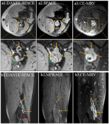
 Figure 1 Representative images from a patient subject. The thrombus-mimicking venous blood signal with the SPACE sequence can be effectively eliminated by DANTE-SPACE (yellow arrows on a1&a2). MPRAGE only detected the DVT in the acute or sub-acute stage because of short T1 relaxation time (yellow arrows on b2), while DANTE-SPACE depicted the DVT well regardless of the thrombus stage (yellow and green arrows on b1) as the venous blood flow (red arrow on b1) around the thrombus was effectively suppressed. The thrombus distribution matched well between DANTE-SPACE and CE-MRV (a1 vs. a3, b1 vs. b3). Table 1 Qualitative and quantitative analysis results for the comparison among DANTE-SPACE, SPACE, MPRAGE, and CE-MRV Score (mean ± std) Number of thrombosed segment SE (%) SP (%) PPV (%) NPV (%) ACC (%) Diagnostic agreement (κ value / p) Interobserver agreement (κ value / p) DANTE-SPACE (reader1)/(reader2) (3.60 ± 0.61)/(3.70 ± 0.46) 22 / 19 90.9 / 94.4 97.8 / 97.9 90.9 / 89.5 97.8 / 98.9 96.4 / 97.3 (0.89 / < 0.01) / (0.90 / < 0.01) 0.73 / <0 .01 SPACE (reader1)/(reader2) (3.22 ± 0.88)/ (2.72 ± 1.02) 20 / 20 86.4 / 83.3 94.4 / 98.9 78.3 / 92.9 95.5 / 94.9 92.0 / 94.6 (0.88 /< 0.01) / (0.75 / < 0.01) 0.63 / <0.01 MPRAGE (reader1)/(reader2) (1.94 ± 0.79)/ (2.61 ± 0.90) 23 / 14 81.8 / 72.2 98.8 / 94.7 95.0 / 75.0 96.7 / 96.7 96.4 / 92.9 (0.75 / < 0.01) / (0.78 / < 0.01) 0.71 / <0.01 CE-MRV (reader1)/(reader2) (3.81 ± 0.42)/ (3.82 ± 0.41) 22 / 18 ---- ---- ---- ---- ---- ---- 0.81 / <0.01 Conclusions DANTE-SPACE is a BTI technique providing excellent venous blood signal suppression and definitive thrombus detection. The preliminary patient study has demonstrated that the technique may outperform SPACE, MPRAGE and potentially become a noncontrast alternative to CE-MRV in the diagnosis of DVT.

          Related collections

          Most cited references5

          • Record: found
          • Abstract: found
          • Article: not found

          DANTE-prepared pulse trains: a novel approach to motion-sensitized and motion-suppressed quantitative magnetic resonance imaging.

          Delay alternating with nutation for tailored excitation (DANTE) pulse trains are well appreciated as frequency-selective excitation methods in Fourier transform NMR and for spatial tagging in MRI. In this study, nonselective DANTE pulse trains are used in combination with gradient pulses and short repetition times as motion-sensitive preparation modules. We show that while the longitudinal magnetization of static tissue is mostly preserved, flowing spins are largely (or fully) attenuated as they fail to establish transverse steady state due to a spoiling effect caused by flow along the applied gradient. The attenuation of flowing spins is effectively insensitive to spin velocity (above a low threshold) and can be approximately quantified with a simple T₁ longitudinal magnetization decay model. The relevant analytical equations for moving spins and static spins during DANTE module application are derived for both transient and steady state epochs. The equations are validated by comparing analytical solutions and numerical Bloch equation simulations against experimental observations in phantoms and in vivo. Based on this contrast mechanism, the application of the DANTE preparation to black blood vessel imaging is proposed. A simple demonstration of DANTE black blood imaging modules shows that it provides excellent blood signal suppression and static tissue signal preservation. Copyright © 2012 Wiley Periodicals, Inc.
            Bookmark
            • Record: found
            • Abstract: found
            • Article: not found

            Three-dimensional black-blood T1-weighted turbo spin-echo techniques for the diagnosis of deep vein thrombosis in comparison with contrast-enhanced magnetic resonance imaging: a pilot study.

            The objective of this study was to evaluate the feasibility of a novel 3-dimensional turbo spin-echo technique with isotropic resolution for the diagnosis of deep vein thrombosis (DVT) in comparison with contrast-enhanced magnetic resonance imaging (CE-MRI) and sonography.
              Bookmark
              • Record: found
              • Abstract: found
              • Article: not found

              Lower-limb deep venous thrombosis: direct MR imaging of the thrombus.

              To determine the feasibility of diagnosing deep venous thrombosis (DVT) with direct three-dimensional magnetic resonance (MR) imaging of the thrombus. Eighteen patients with proved DVT at conventional venography were evaluated with a magnetization-prepared three-dimensional blood- and fat-suppressed MR imaging sequence. The presence and extent of thrombosis on the MR images were compared with the findings at conventional venography. At MR imaging, DVT was visualized in 17 of the 18 patients. MR imaging demonstrated greater proximal extent of the thrombosis in four patients, asymptomatic contralateral thrombosis in one, involvement of the deep femoral vein in five, and involvement of the superficial venous system in three. Direct MR imaging of clots appears capable of demonstrating venographically diagnosed DVT. This MR imaging technique is noninvasive, quick, and repeatable and allows a survey of the whole lower-limb venous system. Detection of thrombosis relies on the formation of methemoglobin, which appears to be sufficiently rapid to allow use of this technique in the clinical setting. Changes in the signal from clot over time may allow estimation of the age of the thrombus.
                Bookmark

                Author and article information

                Conference
                J Cardiovasc Magn Reson
                J Cardiovasc Magn Reson
                Journal of Cardiovascular Magnetic Resonance
                BioMed Central (London )
                1097-6647
                1532-429X
                27 January 2016
                27 January 2016
                2016
                : 18
                Issue : Suppl 1 Issue sponsor : Publication of this supplement was funded by the Society for Cardiovascular Magnetic Resonance.
                : Q58
                Affiliations
                [1 ]Department of Radiology, Guangzhou Panyu Central Hospital, Guangzhou, China
                [2 ]grid.458489.c0000000104837922Shenzhen Institutes of Advanced Technology, Shenzhen, China
                [3 ]grid.413259.80000000406323337Xuanwu Hospital, Beijing, China
                [4 ]grid.50956.3f0000000121529905Biomedical Imaging Research Institute, Cedars-Sinai Medical Center, Los Angeles, CA
                [5 ]Siemens Healthcare, Los Angeles, CA USA
                Article
                5394
                10.1186/1532-429X-18-S1-Q58
                5032585
                09936ebb-2084-475d-a642-d865f0a3d45e
                © Chen et al. 2016

                This article is published under license to BioMed Central Ltd. This is an Open Access article distributed under the terms of the Creative Commons Attribution License ( http://creativecommons.org/licenses/by/4.0), which permits unrestricted use, distribution, and reproduction in any medium, provided the original work is properly cited. The Creative Commons Public Domain Dedication waiver ( http://creativecommons.org/publicdomain/zero/1.0/) applies to the data made available in this article, unless otherwise stated.

                19th Annual SCMR Scientific Sessions
                Los Angeles, CA, USA
                27-30 January 2016
                History
                Categories
                Walking Poster Presentation
                Custom metadata
                © The Author(s) 2016

                Cardiovascular Medicine
                Cardiovascular Medicine

                Comments

                Comment on this article